3G6J

C3b in complex with a C3b specific Fab


Experimental Data Snapshot

  • Method: X-RAY DIFFRACTION
  • Resolution: 3.10 Å
  • R-Value Free: 0.282 
  • R-Value Work: 0.214 
  • R-Value Observed: 0.218 

wwPDB Validation   3D Report Full Report


This is version 1.2 of the entry. See complete history


Literature

Structural and Functional Analysis of a C3b-specific Antibody That Selectively Inhibits the Alternative Pathway of Complement

Katschke, K.J.Stawicki, S.Yin, J.Steffek, M.Xi, H.Sturgeon, L.Hass, P.E.Loyet, K.M.Deforge, L.Wu, Y.van Lookeren Campagne, M.Wiesmann, C.

(2009) J Biol Chem 284: 10473-10479

  • DOI: https://doi.org/10.1074/jbc.M809106200
  • Primary Citation of Related Structures:  
    3G6J

  • PubMed Abstract: 

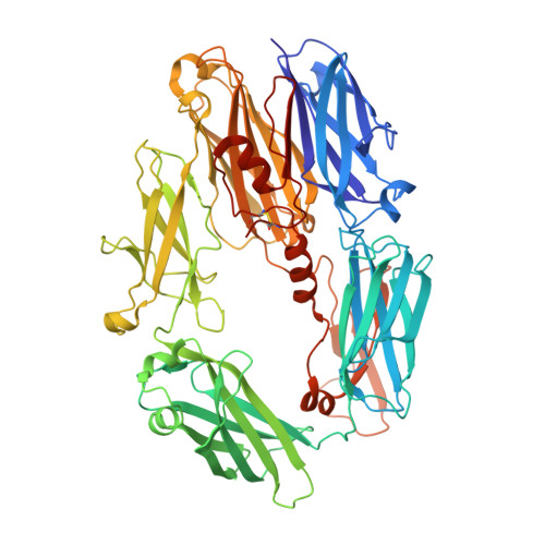
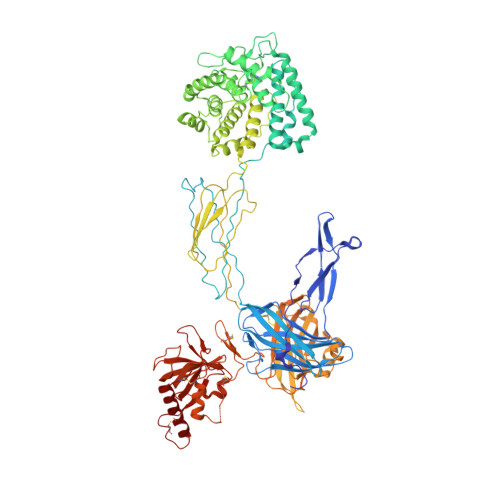
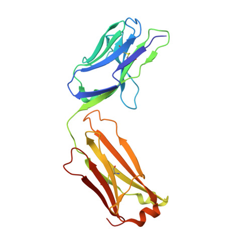
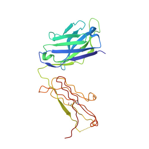
    Amplification of the complement cascade through the alternative pathway can lead to excessive inflammation. Targeting C3b, a component central to the alternative pathway of complement, provides a powerful approach to inhibit complement-mediated immune responses and tissue injury. In the present study, phage display technology was employed to generate an antibody that selectively recognizes C3b but not the non-activated molecule C3. The crystal structure of C3b in complex with a Fab fragment of this antibody (S77) illustrates the structural basis for this selectivity. Cleavage of C3 to C3b results in a plethora of structural changes within C3, including the rearrangement of macroglobulin domain 6 enabling binding of S77 to the adjacent macroglobulin domain 7 domain. S77 blocks binding of factor B to C3b inhibiting the first step in the formation of the alternative pathway C3 convertase. In addition, S77 inhibits C5 binding to C3b. This results in significantly reduced formations of anaphylatoxins and membrane-attack complexes. This study for the first time demonstrates the structural basis for complement inhibition by a C3b-selective antibody and provides insights into the molecular mechanisms of alternative pathway complement activation.


  • Organizational Affiliation

    Departments of Immunology, Antibody Engineering, Protein Engineering, Assay & Automation Technology, and Protein Chemistry, Genentech Inc., 1 DNA Way, South San Francisco, California 94080, USA.


Macromolecules
Find similar proteins by:  (by identity cutoff)  |  3D Structure
Entity ID: 1
MoleculeChains Sequence LengthOrganismDetailsImage
Complement C3 beta chain
A, C
644Homo sapiensMutation(s): 0 
UniProt & NIH Common Fund Data Resources
Find proteins for P01024 (Homo sapiens)
Explore P01024 
Go to UniProtKB:  P01024
PHAROS:  P01024
GTEx:  ENSG00000125730 
Entity Groups  
Sequence Clusters30% Identity50% Identity70% Identity90% Identity95% Identity100% Identity
UniProt GroupP01024
Sequence Annotations
Expand
  • Reference Sequence
Find similar proteins by:  (by identity cutoff)  |  3D Structure
Entity ID: 2
MoleculeChains Sequence LengthOrganismDetailsImage
Complement C3 alpha chain
B, D
915Homo sapiensMutation(s): 0 
UniProt & NIH Common Fund Data Resources
Find proteins for P01024 (Homo sapiens)
Explore P01024 
Go to UniProtKB:  P01024
PHAROS:  P01024
GTEx:  ENSG00000125730 
Entity Groups  
Sequence Clusters30% Identity50% Identity70% Identity90% Identity95% Identity100% Identity
UniProt GroupP01024
Sequence Annotations
Expand
  • Reference Sequence
Find similar proteins by:  (by identity cutoff)  |  3D Structure
Entity ID: 3
MoleculeChains Sequence LengthOrganismDetailsImage
Fab light chain
E, G
214Homo sapiensMutation(s): 0 
Entity Groups  
Sequence Clusters30% Identity50% Identity70% Identity90% Identity95% Identity100% Identity
Sequence Annotations
Expand
  • Reference Sequence
Find similar proteins by:  (by identity cutoff)  |  3D Structure
Entity ID: 4
MoleculeChains Sequence LengthOrganismDetailsImage
Fab heavy chain
F, H
226Homo sapiensMutation(s): 0 
Entity Groups  
Sequence Clusters30% Identity50% Identity70% Identity90% Identity95% Identity100% Identity
Sequence Annotations
Expand
  • Reference Sequence
Experimental Data & Validation

Experimental Data

  • Method: X-RAY DIFFRACTION
  • Resolution: 3.10 Å
  • R-Value Free: 0.282 
  • R-Value Work: 0.214 
  • R-Value Observed: 0.218 
  • Space Group: C 1 2 1
Unit Cell:
Length ( Å )Angle ( ˚ )
a = 216.436α = 90
b = 180.417β = 115.73
c = 154.584γ = 90
Software Package:
Software NamePurpose
HKL-2000data collection
PHASERphasing
REFMACrefinement
HKL-2000data reduction
SCALEPACKdata scaling

Structure Validation

View Full Validation Report


Entry History 

Deposition Data

  • Released Date: 2009-03-10 
  • Deposition Author(s): Wiesmann, C.

Revision History  (Full details and data files)

  • Version 1.0: 2009-03-10
    Type: Initial release
  • Version 1.1: 2011-07-13
    Changes: Advisory, Refinement description, Version format compliance
  • Version 1.2: 2023-09-06
    Changes: Data collection, Database references, Derived calculations, Refinement description