3FWK

Crystal Structure of Candida glabrata FMN Adenylyltransferase


Experimental Data Snapshot

  • Method: X-RAY DIFFRACTION
  • Resolution: 1.20 Å
  • R-Value Free: 0.178 
  • R-Value Work: 0.162 
  • R-Value Observed: 0.162 

wwPDB Validation   3D Report Full Report


Ligand Structure Quality Assessment 


This is version 1.4 of the entry. See complete history


Literature

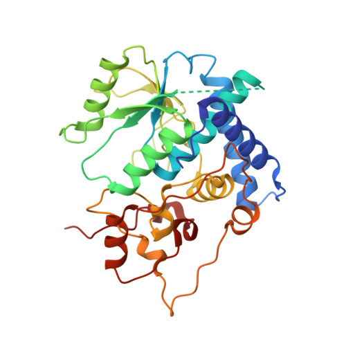
Structure and mechanism of a eukaryotic FMN adenylyltransferase.

Huerta, C.Borek, D.Machius, M.Grishin, N.V.Zhang, H.

(2009) J Mol Biol 389: 388-400

  • DOI: https://doi.org/10.1016/j.jmb.2009.04.022
  • Primary Citation of Related Structures:  
    3FWK, 3G59, 3G5A, 3G6K

  • PubMed Abstract: 

    Flavin mononucleotide adenylyltransferase (FMNAT) catalyzes the formation of the essential flavocoenzyme flavin adenine dinucleotide (FAD) and plays an important role in flavocoenzyme homeostasis regulation. By sequence comparison, bacterial and eukaryotic FMNAT enzymes belong to two different protein superfamilies and apparently utilize different sets of active-site residues to accomplish the same chemistry. Here we report the first structural characterization of a eukaryotic FMNAT from the pathogenic yeast Candida glabrata. Four crystal structures of C. glabrata FMNAT in different complexed forms were determined at 1.20-1.95 A resolutions, capturing the enzyme active-site states prior to and after catalysis. These structures reveal a novel flavin-binding mode and a unique enzyme-bound FAD conformation. Comparison of the bacterial and eukaryotic FMNATs provides a structural basis for understanding the convergent evolution of the same FMNAT activity from different protein ancestors. Structure-based investigation of the kinetic properties of FMNAT should offer insights into the regulatory mechanisms of FAD homeostasis by FMNAT in eukaryotic organisms.


  • Organizational Affiliation

    Department of Biochemistry, University of Texas Southwestern Medical Center, Dallas, TX 75390, USA.


Macromolecules
Find similar proteins by:  (by identity cutoff)  |  3D Structure
Entity ID: 1
MoleculeChains Sequence LengthOrganismDetailsImage
FMN Adenylyltransferase308Nakaseomyces glabratusMutation(s): 0 
Gene Names: CAGL0K01397gFAD1
EC: 2.7.7.2
UniProt
Find proteins for Q6FNA9 (Candida glabrata (strain ATCC 2001 / BCRC 20586 / JCM 3761 / NBRC 0622 / NRRL Y-65 / CBS 138))
Explore Q6FNA9 
Go to UniProtKB:  Q6FNA9
Entity Groups  
Sequence Clusters30% Identity50% Identity70% Identity90% Identity95% Identity100% Identity
UniProt GroupQ6FNA9
Sequence Annotations
Expand
  • Reference Sequence
Small Molecules
Experimental Data & Validation

Experimental Data

  • Method: X-RAY DIFFRACTION
  • Resolution: 1.20 Å
  • R-Value Free: 0.178 
  • R-Value Work: 0.162 
  • R-Value Observed: 0.162 
  • Space Group: P 32 2 1
Unit Cell:
Length ( Å )Angle ( ˚ )
a = 80.09α = 90
b = 80.09β = 90
c = 78.089γ = 120
Software Package:
Software NamePurpose
DENZOdata reduction
SCALEPACKdata scaling
REFMACrefinement
PDB_EXTRACTdata extraction
HKL-3000data collection
HKL-3000data reduction
HKL-3000data scaling
REFMACphasing

Structure Validation

View Full Validation Report


Ligand Structure Quality Assessment 


Entry History 

Deposition Data

Revision History  (Full details and data files)

  • Version 1.0: 2009-05-26
    Type: Initial release
  • Version 1.1: 2011-07-13
    Changes: Version format compliance
  • Version 1.2: 2017-11-01
    Changes: Refinement description
  • Version 1.3: 2020-07-29
    Type: Remediation
    Reason: Carbohydrate remediation
    Changes: Data collection, Database references, Derived calculations, Structure summary
  • Version 1.4: 2024-02-21
    Changes: Data collection, Database references, Structure summary