3FVX

Human kynurenine aminotransferase I in complex with tris


Experimental Data Snapshot

  • Method: X-RAY DIFFRACTION
  • Resolution: 1.50 Å
  • R-Value Free: 0.200 
  • R-Value Work: 0.178 
  • R-Value Observed: 0.180 

wwPDB Validation   3D Report Full Report


This is version 1.3 of the entry. See complete history


Literature

Structural insight into the inhibition of human kynurenine aminotransferase I/glutamine transaminase K

Han, Q.Robinson, H.Cai, T.Tagle, D.A.Li, J.

(2009) J Med Chem 52: 2786-2793

  • DOI: https://doi.org/10.1021/jm9000874
  • Primary Citation of Related Structures:  
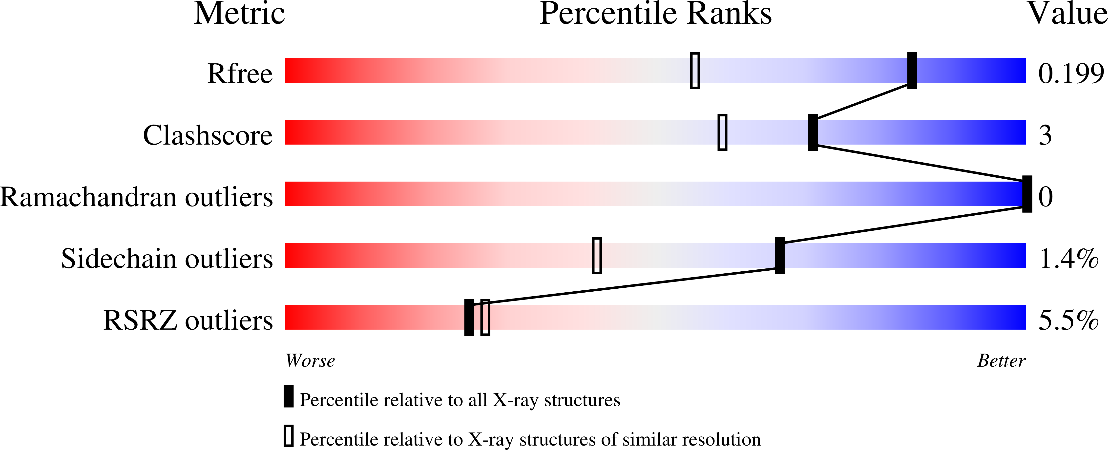
    3FVS, 3FVU, 3FVX

  • PubMed Abstract: 

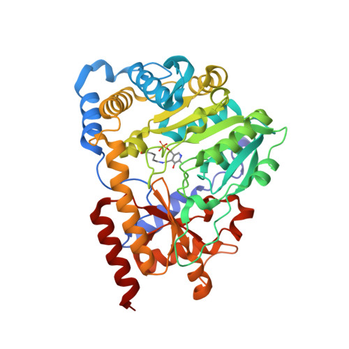
    Human kynurenine aminotransferase I (hKAT I) catalyzes the formation of kynurenic acid, a neuroactive compound. Here, we report three high-resolution crystal structures (1.50-1.55 A) of hKAT I that are in complex with glycerol and each of two inhibitors of hKAT I: indole-3-acetic acid (IAC) and Tris. Because Tris is able to occupy the substrate binding position, we speculate that this may be the basis for hKAT I inhibition. Furthermore, the hKAT/IAC complex structure reveals that the binding moieties of the inhibitor are its indole ring and a carboxyl group. Six chemicals with both binding moieties were tested for their ability to inhibit hKAT I activity; 3-indolepropionic acid and DL-indole-3-lactic acid demonstrated the highest level of inhibition, and as they cannot be considered as substrates of the enzyme, these two inhibitors are promising candidates for future study. Perhaps even more significantly, we report the discovery of two different ligands located simultaneously in the hKAT I active center for the first time.


  • Organizational Affiliation

    Department of Biochemistry, Virginia Tech, Blacksburg, Virginia 24061, USA. qianhan@vt.edu


Macromolecules
Find similar proteins by:  (by identity cutoff)  |  3D Structure
Entity ID: 1
MoleculeChains Sequence LengthOrganismDetailsImage
Kynurenine--oxoglutarate transaminase 1
A, B
422Homo sapiensMutation(s): 0 
Gene Names: CCBL1
EC: 2.6.1.7 (PDB Primary Data), 2.6.1.64 (PDB Primary Data), 4.4.1.13 (PDB Primary Data)
UniProt & NIH Common Fund Data Resources
Find proteins for Q16773 (Homo sapiens)
Explore Q16773 
Go to UniProtKB:  Q16773
PHAROS:  Q16773
GTEx:  ENSG00000171097 
Entity Groups  
Sequence Clusters30% Identity50% Identity70% Identity90% Identity95% Identity100% Identity
UniProt GroupQ16773
Sequence Annotations
Expand
  • Reference Sequence
Small Molecules
Modified Residues  1 Unique
IDChains TypeFormula2D DiagramParent
LLP
Query on LLP
A, B
L-PEPTIDE LINKINGC14 H22 N3 O7 PLYS
Experimental Data & Validation

Experimental Data

  • Method: X-RAY DIFFRACTION
  • Resolution: 1.50 Å
  • R-Value Free: 0.200 
  • R-Value Work: 0.178 
  • R-Value Observed: 0.180 
  • Space Group: C 1 2 1
Unit Cell:
Length ( Å )Angle ( ˚ )
a = 102.881α = 90
b = 107.439β = 112.87
c = 81.686γ = 90
Software Package:
Software NamePurpose
HKL-2000data collection
MOLREPphasing
REFMACrefinement
DENZOdata reduction
SCALAdata scaling

Structure Validation

View Full Validation Report


Entry History 

Deposition Data

Revision History  (Full details and data files)

  • Version 1.0: 2009-05-19
    Type: Initial release
  • Version 1.1: 2011-07-13
    Changes: Version format compliance
  • Version 1.2: 2023-09-06
    Changes: Data collection, Database references, Derived calculations, Refinement description
  • Version 1.3: 2023-11-22
    Changes: Data collection