3FQT

Phosphorylation of self-peptides alters Human Leukocyte Antigen Class I-restricted antigen presentation and generates tumor specific epitopes


Experimental Data Snapshot

  • Method: X-RAY DIFFRACTION
  • Resolution: 1.80 Å
  • R-Value Free: 0.204 
  • R-Value Work: 0.174 
  • R-Value Observed: 0.176 

wwPDB Validation   3D Report Full Report


This is version 1.3 of the entry. See complete history


Literature

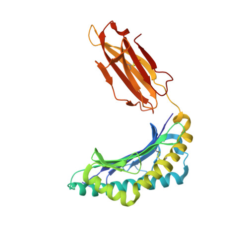
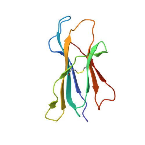
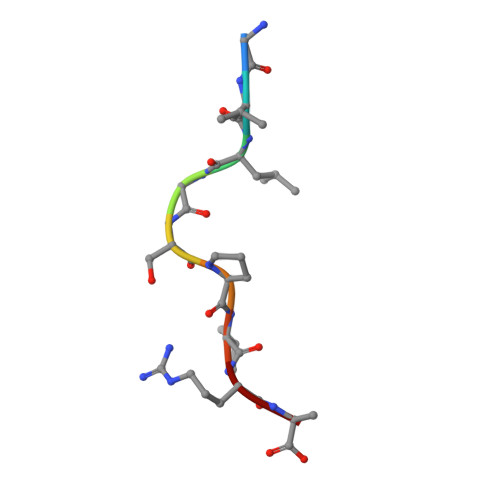
Phosphorylated self-peptides alter human leukocyte antigen class I-restricted antigen presentation and generate tumor-specific epitopes

Petersen, J.Wurzbacher, S.J.Williamson, N.A.Ramarathinam, S.H.Reid, H.H.Nair, A.K.Zhao, A.Y.Nastovska, R.Rudge, G.Rossjohn, J.Purcell, A.W.

(2009) Proc Natl Acad Sci U S A 106: 2776-2781

  • DOI: https://doi.org/10.1073/pnas.0812901106
  • Primary Citation of Related Structures:  
    3FQN, 3FQR, 3FQT, 3FQU, 3FQW, 3FQX

  • PubMed Abstract: 

    Human leukocyte antigen (HLA) class I molecules present a variety of posttranslationally modified epitopes at the cell surface, although the consequences of such presentation remain largely unclear. Phosphorylation plays a critical cellular role, and deregulation in phosphate metabolism is associated with disease, including autoimmunity and tumor immunity. We have solved the high-resolution structures of 3 HLA A2-restricted phosphopeptides associated with tumor immunity and compared them with the structures of their nonphosphorylated counterparts. Phosphorylation of the epitope was observed to affect the structure and mobility of the bound epitope. In addition, the phosphoamino acid stabilized the HLA peptide complex in an epitope-specific manner and was observed to exhibit discrete flexibility within the antigen-binding cleft. Collectively, our data suggest that phosphorylation generates neoepitopes that represent demanding targets for T-cell receptor ligation. These findings provide insights into the mode of phosphopeptide presentation by HLA as well as providing a platform for the rational design of a generation of posttranslationally modified tumor vaccines.


  • Organizational Affiliation

    Protein Crystallography Unit, Department of Biochemistry and Molecular Biology, School of Biomedical Sciences, Monash University, Victoria 3800, Australia.


Macromolecules
Find similar proteins by:  (by identity cutoff)  |  3D Structure
Entity ID: 1
MoleculeChains Sequence LengthOrganismDetailsImage
HLA class I histocompatibility antigen, A-2 alpha chain275Homo sapiensMutation(s): 0 
Gene Names: HLA-AHLAA
UniProt & NIH Common Fund Data Resources
Find proteins for P04439 (Homo sapiens)
Explore P04439 
Go to UniProtKB:  P04439
PHAROS:  P04439
GTEx:  ENSG00000206503 
Entity Groups  
Sequence Clusters30% Identity50% Identity70% Identity90% Identity95% Identity100% Identity
UniProt GroupP04439
Sequence Annotations
Expand
  • Reference Sequence
Find similar proteins by:  (by identity cutoff)  |  3D Structure
Entity ID: 2
MoleculeChains Sequence LengthOrganismDetailsImage
Beta-2-microglobulin98Homo sapiensMutation(s): 0 
Gene Names: B2M
UniProt & NIH Common Fund Data Resources
Find proteins for P61769 (Homo sapiens)
Explore P61769 
Go to UniProtKB:  P61769
PHAROS:  P61769
GTEx:  ENSG00000166710 
Entity Groups  
Sequence Clusters30% Identity50% Identity70% Identity90% Identity95% Identity100% Identity
UniProt GroupP61769
Sequence Annotations
Expand
  • Reference Sequence

Find similar proteins by:  Sequence   |   3D Structure  

Entity ID: 3
MoleculeChains Sequence LengthOrganismDetailsImage
peptide 38-46 from cell division cycle 25b (CDC25b): GLLGSPVRA9N/AMutation(s): 0 
UniProt & NIH Common Fund Data Resources
Find proteins for P30305 (Homo sapiens)
Explore P30305 
Go to UniProtKB:  P30305
PHAROS:  P30305
GTEx:  ENSG00000101224 
Entity Groups  
UniProt GroupP30305
Sequence Annotations
Expand
  • Reference Sequence
Small Molecules
Ligands 3 Unique
IDChains Name / Formula / InChI Key2D Diagram3D Interactions
CD
Query on CD

Download Ideal Coordinates CCD File 
D [auth A]
E [auth A]
F [auth A]
G [auth A]
H [auth A]
D [auth A],
E [auth A],
F [auth A],
G [auth A],
H [auth A],
N [auth B]
CADMIUM ION
Cd
WLZRMCYVCSSEQC-UHFFFAOYSA-N
GOL
Query on GOL

Download Ideal Coordinates CCD File 
J [auth A]
K [auth A]
L [auth A]
M [auth A]
O [auth B]
J [auth A],
K [auth A],
L [auth A],
M [auth A],
O [auth B],
P [auth C]
GLYCEROL
C3 H8 O3
PEDCQBHIVMGVHV-UHFFFAOYSA-N
MG
Query on MG

Download Ideal Coordinates CCD File 
I [auth A]MAGNESIUM ION
Mg
JLVVSXFLKOJNIY-UHFFFAOYSA-N
Experimental Data & Validation

Experimental Data

  • Method: X-RAY DIFFRACTION
  • Resolution: 1.80 Å
  • R-Value Free: 0.204 
  • R-Value Work: 0.174 
  • R-Value Observed: 0.176 
  • Space Group: P 21 21 21
Unit Cell:
Length ( Å )Angle ( ˚ )
a = 59.68α = 90
b = 79.84β = 90
c = 110.207γ = 90
Software Package:
Software NamePurpose
DENZOdata reduction
SCALEPACKdata scaling
PHENIXrefinement
PDB_EXTRACTdata extraction
CrystalCleardata collection
HKL-2000data reduction
PHASERphasing

Structure Validation

View Full Validation Report


Entry History 

Deposition Data

Revision History  (Full details and data files)

  • Version 1.0: 2009-03-03
    Type: Initial release
  • Version 1.1: 2011-07-13
    Changes: Non-polymer description, Version format compliance
  • Version 1.2: 2017-11-01
    Changes: Advisory, Refinement description
  • Version 1.3: 2023-11-01
    Changes: Advisory, Data collection, Database references, Derived calculations, Refinement description