3FP8

Anionic trypsin variant S195A in complex with bovine pancreatic trypsin inhibitor (BPTI) determined to the 1.46 A resolution limit


Experimental Data Snapshot

  • Method: X-RAY DIFFRACTION
  • Resolution: 1.46 Å
  • R-Value Free: 0.189 
  • R-Value Work: 0.175 
  • R-Value Observed: 0.175 

wwPDB Validation   3D Report Full Report


This is version 1.5 of the entry. See complete history


Literature

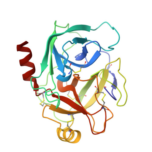
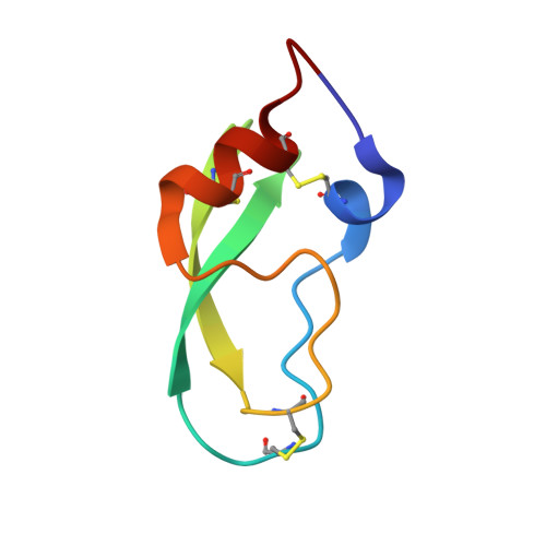
Structure of a serine protease poised to resynthesize a peptide bond.

Zakharova, E.Horvath, M.P.Goldenberg, D.P.

(2009) Proc Natl Acad Sci U S A 106: 11034-11039

  • DOI: https://doi.org/10.1073/pnas.0902463106
  • Primary Citation of Related Structures:  
    3FP6, 3FP7, 3FP8

  • PubMed Abstract: 

    The serine proteases are among the most thoroughly studied enzymes, and numerous crystal structures representing the enzyme-substrate complex and intermediates in the hydrolysis reactions have been reported. Some aspects of the catalytic mechanism remain controversial, however, especially the role of conformational changes in the reaction. We describe here a high-resolution (1.46 A) crystal structure of a complex formed between a cleaved form of bovine pancreatic trypsin inhibitor (BPTI) and a catalytically inactive trypsin variant with the BPTI cleavage site ideally positioned in the active site for resynthesis of the peptide bond. This structure defines the positions of the newly generated amino and carboxyl groups following the 2 steps in the hydrolytic reaction. Comparison of this structure with those representing other intermediates in the reaction demonstrates that the residues of the catalytic triad are positioned to promote each step of both the forward and reverse reaction with remarkably little motion and with conservation of hydrogen-bonding interactions. The results also provide insights into the mechanism by which inhibitors like BPTI normally resist hydrolysis when bound to their target proteases.


  • Organizational Affiliation

    Department of Biology, University of Utah, Salt Lake City, UT 84112-0840, USA.


Macromolecules
Find similar proteins by:  (by identity cutoff)  |  3D Structure
Entity ID: 1
MoleculeChains Sequence LengthOrganismDetailsImage
Anionic trypsin-2A [auth E]223Rattus norvegicusMutation(s): 1 
Gene Names: Prss2Try2
EC: 3.4.21.4
UniProt
Find proteins for P00763 (Rattus norvegicus)
Explore P00763 
Go to UniProtKB:  P00763
Entity Groups  
Sequence Clusters30% Identity50% Identity70% Identity90% Identity95% Identity100% Identity
UniProt GroupP00763
Sequence Annotations
Expand
  • Reference Sequence
Find similar proteins by:  (by identity cutoff)  |  3D Structure
Entity ID: 2
MoleculeChains Sequence LengthOrganismDetailsImage
Pancreatic trypsin inhibitorB [auth I]58Bos taurusMutation(s): 0 
UniProt
Find proteins for P00974 (Bos taurus)
Explore P00974 
Go to UniProtKB:  P00974
Entity Groups  
Sequence Clusters30% Identity50% Identity70% Identity90% Identity95% Identity100% Identity
UniProt GroupP00974
Sequence Annotations
Expand
  • Reference Sequence
Small Molecules
Ligands 4 Unique
IDChains Name / Formula / InChI Key2D Diagram3D Interactions
PG4
Query on PG4

Download Ideal Coordinates CCD File 
H [auth E]TETRAETHYLENE GLYCOL
C8 H18 O5
UWHCKJMYHZGTIT-UHFFFAOYSA-N
SO4
Query on SO4

Download Ideal Coordinates CCD File 
J [auth E],
L [auth I],
M [auth I],
N [auth I]
SULFATE ION
O4 S
QAOWNCQODCNURD-UHFFFAOYSA-L
EDO
Query on EDO

Download Ideal Coordinates CCD File 
C [auth E]
D [auth E]
E
F [auth E]
G [auth E]
C [auth E],
D [auth E],
E,
F [auth E],
G [auth E],
K [auth I]
1,2-ETHANEDIOL
C2 H6 O2
LYCAIKOWRPUZTN-UHFFFAOYSA-N
CA
Query on CA

Download Ideal Coordinates CCD File 
I [auth E]CALCIUM ION
Ca
BHPQYMZQTOCNFJ-UHFFFAOYSA-N
Experimental Data & Validation

Experimental Data

  • Method: X-RAY DIFFRACTION
  • Resolution: 1.46 Å
  • R-Value Free: 0.189 
  • R-Value Work: 0.175 
  • R-Value Observed: 0.175 
  • Space Group: P 32 2 1
Unit Cell:
Length ( Å )Angle ( ˚ )
a = 91.933α = 90
b = 91.933β = 90
c = 61.135γ = 120
Software Package:
Software NamePurpose
SBC-Collectdata collection
CNSrefinement
HKL-2000data reduction
HKL-2000data scaling
CNSphasing

Structure Validation

View Full Validation Report


Entry History 

Deposition Data

Revision History  (Full details and data files)

  • Version 1.0: 2009-02-17
    Type: Initial release
  • Version 1.1: 2011-07-13
    Changes: Version format compliance
  • Version 1.2: 2017-11-01
    Changes: Refinement description
  • Version 1.3: 2018-04-04
    Changes: Data collection
  • Version 1.4: 2021-10-20
    Changes: Database references, Derived calculations
  • Version 1.5: 2023-09-06
    Changes: Data collection, Refinement description