3FHR

High resolution crystal structure of mitogen-activated protein kinase-activated protein kinase 3 (MK3)-inhibitor complex


Experimental Data Snapshot

  • Method: X-RAY DIFFRACTION
  • Resolution: 1.90 Å
  • R-Value Free: 0.267 
  • R-Value Work: 0.226 
  • R-Value Observed: 0.228 

wwPDB Validation   3D Report Full Report


Ligand Structure Quality Assessment 


This is version 1.2 of the entry. See complete history

Re-refinement Note

A newer entry is available that reflects an alternative modeling of the original data: 7NRB 7NRY


Literature

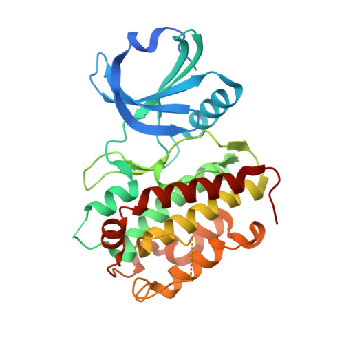
High-resolution crystal structure of human Mapkap kinase 3 in complex with a high affinity ligand

Cheng, R.Felicetti, B.Palan, S.Toogood-Johnson, I.Scheich, C.Barker, J.Whittaker, M.Hesterkamp, T.

(2010) Protein Sci 19: 168-173

  • DOI: https://doi.org/10.1002/pro.294
  • Primary Citation of Related Structures:  
    3FHR

  • PubMed Abstract: 

    The Mapkap kinases 2 and 3 (MK2 and MK3) have been implicated in intracellular signaling pathways leading to the production of the pro-inflammatory cytokine tumor necrosis factor alpha. MK2 has been pursued by the biopharmaceutical industry for many years for the development of a small molecule anti-inflammatory treatment and drug-like inhibitors have been described. The development of some of these compounds, however, has been slowed by the absence of a high-resolution crystal structure of MK2. Herein we present a high-resolution (1.9 A) crystal structure of the highly homologous MK3 in complex with a pharmaceutical lead compound. While all of the canonical features of Ser/Thr kinases in general and MK2 in particular are recapitulated in MK3, the detailed analysis of the binding interaction of the drug-like ligand within the adenine binding pocket allows relevant conclusions to be drawn for the further design of potent and selective drug candidates.


  • Organizational Affiliation

    Evotec (UK) Ltd, 114 Milton Park, Abingdon, Oxfordshire OX14 4SA, United Kingdom.


Macromolecules
Find similar proteins by:  (by identity cutoff)  |  3D Structure
Entity ID: 1
MoleculeChains Sequence LengthOrganismDetailsImage
MAP kinase-activated protein kinase 3336Homo sapiensMutation(s): 0 
EC: 2.7.11.1
UniProt & NIH Common Fund Data Resources
Find proteins for Q16644 (Homo sapiens)
Explore Q16644 
Go to UniProtKB:  Q16644
PHAROS:  Q16644
GTEx:  ENSG00000114738 
Entity Groups  
Sequence Clusters30% Identity50% Identity70% Identity90% Identity95% Identity100% Identity
UniProt GroupQ16644
Sequence Annotations
Expand
  • Reference Sequence
Small Molecules
Ligands 1 Unique
IDChains Name / Formula / InChI Key2D Diagram3D Interactions
P4O
Query on P4O

Download Ideal Coordinates CCD File 
B [auth A]2-(2-QUINOLIN-3-YLPYRIDIN-4-YL)-1,5,6,7-TETRAHYDRO-4H-PYRROLO[3,2-C]PYRIDIN-4-ONE
C21 H16 N4 O
OWFLADWRSCINST-UHFFFAOYSA-N
Binding Affinity Annotations 
IDSourceBinding Affinity
P4O BindingDB:  3FHR IC50: 210 (nM) from 1 assay(s)
PDBBind:  3FHR IC50: 210 (nM) from 1 assay(s)
Experimental Data & Validation

Experimental Data

  • Method: X-RAY DIFFRACTION
  • Resolution: 1.90 Å
  • R-Value Free: 0.267 
  • R-Value Work: 0.226 
  • R-Value Observed: 0.228 
  • Space Group: C 1 2 1
Unit Cell:
Length ( Å )Angle ( ˚ )
a = 84.688α = 90
b = 74.832β = 107.34
c = 60.664γ = 90
Software Package:
Software NamePurpose
DNAdata collection
PHASERphasing
REFMACrefinement
CrystalCleardata reduction
CrystalCleardata scaling

Structure Validation

View Full Validation Report


Ligand Structure Quality Assessment 


Entry History 

Deposition Data

Revision History  (Full details and data files)

  • Version 1.0: 2009-12-15
    Type: Initial release
  • Version 1.1: 2011-07-13
    Changes: Version format compliance
  • Version 1.2: 2014-01-29
    Changes: Database references