3FAQ

Crystal structure of lactoperoxidase complex with cyanide


Experimental Data Snapshot

  • Method: X-RAY DIFFRACTION
  • Resolution: 2.70 Å
  • R-Value Free: 0.235 
  • R-Value Work: 0.208 
  • R-Value Observed: 0.208 

wwPDB Validation   3D Report Full Report


Ligand Structure Quality Assessment 


This is version 3.1 of the entry. See complete history


Literature

Structural Evidence of Substrate Specificity in Mammalian Peroxidases: STRUCTURE OF THE THIOCYANATE COMPLEX WITH LACTOPEROXIDASE AND ITS INTERACTIONS AT 2.4 A RESOLUTION

Sheikh, I.A.Singh, A.K.Singh, N.Sinha, M.Singh, S.B.Bhushan, A.Kaur, P.Srinivasan, A.Sharma, S.Singh, T.P.

(2009) J Biol Chem 284: 14849-14856

  • DOI: https://doi.org/10.1074/jbc.M807644200
  • Primary Citation of Related Structures:  
    3ERH, 3ERI, 3FAQ

  • PubMed Abstract: 

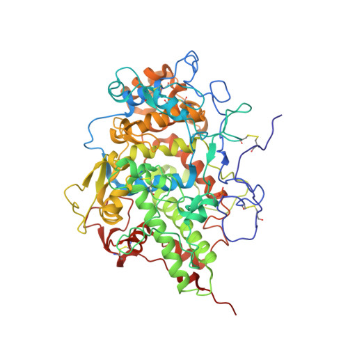
    The crystal structure of the complex of lactoperoxidase (LPO) with its physiological substrate thiocyanate (SCN(-)) has been determined at 2.4A resolution. It revealed that the SCN(-) ion is bound to LPO in the distal heme cavity. The observed orientation of the SCN(-) ion shows that the sulfur atom is closer to the heme iron than the nitrogen atom. The nitrogen atom of SCN(-) forms a hydrogen bond with a water (Wat) molecule at position 6'. This water molecule is stabilized by two hydrogen bonds with Gln(423) N(epsilon2) and Phe(422) oxygen. In contrast, the placement of the SCN(-) ion in the structure of myeloperoxidase (MPO) occurs with an opposite orientation, in which the nitrogen atom is closer to the heme iron than the sulfur atom. The site corresponding to the positions of Gln(423), Phe(422) oxygen, and Wat(6)' in LPO is occupied primarily by the side chain of Phe(407) in MPO due to an entirely different conformation of the loop corresponding to the segment Arg(418)-Phe(431) of LPO. This arrangement in MPO does not favor a similar orientation of the SCN(-) ion. The orientation of the catalytic product OSCN(-) as reported in the structure of LPO.OSCN(-) is similar to the orientation of SCN(-) in the structure of LPO.SCN(-). Similarly, in the structure of LPO.SCN(-).CN(-), in which CN(-) binds at Wat(1), the position and orientation of the SCN(-) ion are also identical to that observed in the structure of LPO.SCN.


  • Organizational Affiliation

    Department of Biophysics, All India Institute of Medical Sciences, New Delhi 110 029, India.


Macromolecules
Find similar proteins by:  (by identity cutoff)  |  3D Structure
Entity ID: 1
MoleculeChains Sequence LengthOrganismDetailsImage
Lactoperoxidase595Bubalus bubalisMutation(s): 0 
EC: 1.11.1.7
UniProt
Find proteins for A5JUY8 (Bubalus bubalis)
Explore A5JUY8 
Go to UniProtKB:  A5JUY8
Entity Groups  
Sequence Clusters30% Identity50% Identity70% Identity90% Identity95% Identity100% Identity
UniProt GroupA5JUY8
Sequence Annotations
Expand
  • Reference Sequence
Oligosaccharides

Help

Entity ID: 2
MoleculeChains Length2D Diagram Glycosylation3D Interactions
2-acetamido-2-deoxy-beta-D-glucopyranose-(1-4)-2-acetamido-2-deoxy-beta-D-glucopyranose
B, C
2N-Glycosylation
Glycosylation Resources
GlyTouCan:  G42666HT
GlyCosmos:  G42666HT
GlyGen:  G42666HT
Entity ID: 3
MoleculeChains Length2D Diagram Glycosylation3D Interactions
alpha-D-mannopyranose-(1-4)-2-acetamido-2-deoxy-beta-D-glucopyranose-(1-4)-2-acetamido-2-deoxy-beta-D-glucopyranose
D
3N-Glycosylation
Glycosylation Resources
GlyTouCan:  G62182OO
GlyCosmos:  G62182OO
GlyGen:  G62182OO
Entity ID: 4
MoleculeChains Length2D Diagram Glycosylation3D Interactions
alpha-D-mannopyranose-(1-4)-beta-D-mannopyranose-(1-4)-2-acetamido-2-deoxy-beta-D-glucopyranose-(1-4)-2-acetamido-2-deoxy-beta-D-glucopyranose
E
4N-Glycosylation
Glycosylation Resources
GlyTouCan:  G08918EL
GlyCosmos:  G08918EL
GlyGen:  G08918EL
Small Molecules
Ligands 5 Unique
IDChains Name / Formula / InChI Key2D Diagram3D Interactions
HEM
Query on HEM

Download Ideal Coordinates CCD File 
F [auth A]PROTOPORPHYRIN IX CONTAINING FE
C34 H32 Fe N4 O4
KABFMIBPWCXCRK-RGGAHWMASA-L
IOD
Query on IOD

Download Ideal Coordinates CCD File 
K [auth A]
L [auth A]
M [auth A]
N [auth A]
O [auth A]
K [auth A],
L [auth A],
M [auth A],
N [auth A],
O [auth A],
P [auth A],
Q [auth A]
IODIDE ION
I
XMBWDFGMSWQBCA-UHFFFAOYSA-M
SCN
Query on SCN

Download Ideal Coordinates CCD File 
G [auth A],
H [auth A]
THIOCYANATE ION
C N S
ZMZDMBWJUHKJPS-UHFFFAOYSA-M
CA
Query on CA

Download Ideal Coordinates CCD File 
I [auth A]CALCIUM ION
Ca
BHPQYMZQTOCNFJ-UHFFFAOYSA-N
CYN
Query on CYN

Download Ideal Coordinates CCD File 
J [auth A]CYANIDE ION
C N
XFXPMWWXUTWYJX-UHFFFAOYSA-N
Modified Residues  1 Unique
IDChains TypeFormula2D DiagramParent
SEP
Query on SEP
A
L-PEPTIDE LINKINGC3 H8 N O6 PSER
Experimental Data & Validation

Experimental Data

  • Method: X-RAY DIFFRACTION
  • Resolution: 2.70 Å
  • R-Value Free: 0.235 
  • R-Value Work: 0.208 
  • R-Value Observed: 0.208 
  • Space Group: P 1 21 1
Unit Cell:
Length ( Å )Angle ( ˚ )
a = 54.453α = 90
b = 80.665β = 102.87
c = 77.796γ = 90
Software Package:
Software NamePurpose
CNSrefinement
MAR345dtbdata collection
AMoREphasing
DENZOdata reduction
SCALEPACKdata scaling

Structure Validation

View Full Validation Report


Ligand Structure Quality Assessment 


Entry History 

Deposition Data

Revision History  (Full details and data files)

  • Version 1.0: 2009-03-31
    Type: Initial release
  • Version 1.1: 2011-07-13
    Changes: Non-polymer description, Version format compliance
  • Version 1.2: 2016-12-21
    Changes: Structure summary
  • Version 2.0: 2020-07-29
    Type: Remediation
    Reason: Carbohydrate remediation
    Changes: Atomic model, Data collection, Derived calculations, Structure summary
  • Version 3.0: 2023-06-28
    Changes: Advisory, Atomic model, Data collection, Database references, Derived calculations, Refinement description, Source and taxonomy, Structure summary
  • Version 3.1: 2023-11-08
    Changes: Data collection, Refinement description