3F3R

Crystal structure of yeast Thioredoxin1-glutathione mixed disulfide complex


Experimental Data Snapshot

  • Method: X-RAY DIFFRACTION
  • Resolution: 1.80 Å
  • R-Value Free: 0.220 
  • R-Value Work: 0.183 
  • R-Value Observed: 0.187 

wwPDB Validation   3D Report Full Report


Ligand Structure Quality Assessment 


This is version 1.4 of the entry. See complete history


Literature

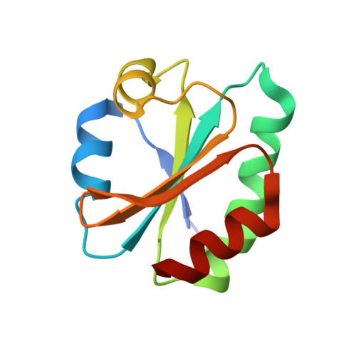
Structural and kinetic analysis of Saccharomyces cerevisiae thioredoxin Trx1: implications for the catalytic mechanism of GSSG reduced by the thioredoxin system

Bao, R.Zhang, Y.R.Lou, X.Zhou, C.Z.Chen, Y.X.

(2009) Biochim Biophys Acta 1794: 1218-1223

  • DOI: https://doi.org/10.1016/j.bbapap.2009.04.001
  • Primary Citation of Related Structures:  
    3F3Q, 3F3R

  • PubMed Abstract: 

    Thioredoxin (Trx) and glutathione/glutaredoxin (GSH/Grx) systems play the dominant role in cellular redox homeostasis. Recently the Trx system has been shown to be responsible to control the balance of GSH/GSSG once the glutathione reductase system is not available. To decipher the structural basis of electron transfer from the Trx system to GSSG, we solved the crystal structures of oxidized Trx1 and glutathionylated Trx1Cys33Ser mutant at 1.76 and 1.80 A, respectively. Comparative structural analysis revealed a key residue Met35 involved in the Trx-GSSG recognition. Subsequent mutagenesis and kinetic studies proved that Met35Arg mutation could alter the apparent K(m) and V(max) values of the reaction. These findings gave us the structural insights into GSSG reduction catalyzed by the Trx system.


  • Organizational Affiliation

    Institute of Protein Research, Tongji University, Shanghai, PR China.


Macromolecules
Find similar proteins by:  (by identity cutoff)  |  3D Structure
Entity ID: 1
MoleculeChains Sequence LengthOrganismDetailsImage
Thioredoxin-1
A, B
109Saccharomyces cerevisiaeMutation(s): 1 
Gene Names: TRX1
UniProt
Find proteins for P22217 (Saccharomyces cerevisiae (strain ATCC 204508 / S288c))
Explore P22217 
Go to UniProtKB:  P22217
Entity Groups  
Sequence Clusters30% Identity50% Identity70% Identity90% Identity95% Identity100% Identity
UniProt GroupP22217
Sequence Annotations
Expand
  • Reference Sequence
Experimental Data & Validation

Experimental Data

  • Method: X-RAY DIFFRACTION
  • Resolution: 1.80 Å
  • R-Value Free: 0.220 
  • R-Value Work: 0.183 
  • R-Value Observed: 0.187 
  • Space Group: P 1
Unit Cell:
Length ( Å )Angle ( ˚ )
a = 38.53α = 72.91
b = 38.81β = 87.51
c = 41.7γ = 60.58
Software Package:
Software NamePurpose
MAR345dtbdata collection
PHASESphasing
REFMACrefinement
MOSFLMdata reduction
SCALAdata scaling

Structure Validation

View Full Validation Report


Ligand Structure Quality Assessment 


Entry History 

Deposition Data

Revision History  (Full details and data files)

  • Version 1.0: 2009-10-20
    Type: Initial release
  • Version 1.1: 2011-07-13
    Changes: Version format compliance
  • Version 1.2: 2012-03-21
    Changes: Non-polymer description
  • Version 1.3: 2021-11-10
    Changes: Database references, Derived calculations
  • Version 1.4: 2023-11-01
    Changes: Data collection, Refinement description