3F2E

Crystal structure of Yellowstone SIRV coat protein C-terminus


Experimental Data Snapshot

  • Method: X-RAY DIFFRACTION
  • Resolution: 1.67 Å
  • R-Value Free: 0.202 
  • R-Value Work: 0.184 
  • R-Value Observed: 0.185 

wwPDB Validation   3D Report Full Report


This is version 1.5 of the entry. See complete history


Literature

Synergy of NMR, computation, and X-ray crystallography for structural biology.

Szymczyna, B.R.Taurog, R.E.Young, M.J.Snyder, J.C.Johnson, J.E.Williamson, J.R.

(2009) Structure 17: 499-507

  • DOI: https://doi.org/10.1016/j.str.2009.03.001
  • Primary Citation of Related Structures:  
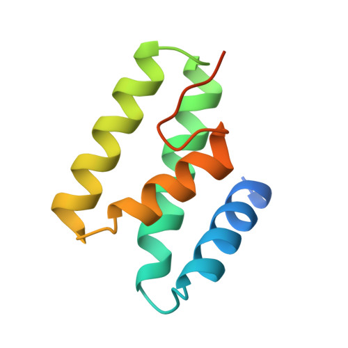
    3F2E

  • PubMed Abstract: 

    NMR spectroscopy and X-ray crystallography are currently the two most widely applied methods for the determination of macromolecular structures at high resolution. More recently, significant advances have been made in algorithms for the de novo prediction of protein structure, and, in favorable cases, the predicted models agree extremely well with experimentally determined structures. Here, we demonstrate a synergistic combination of NMR spectroscopy, de novo structure prediction, and X-ray crystallography in an effective overall strategy for rapidly determining the structure of the coat protein C-terminal domain from the Sulfolobus islandicus rod-shaped virus (SIRV). This approach takes advantage of the most accessible aspects of each structural technique and may be widely applicable for structure determination.


  • Organizational Affiliation

    Department of Molecular Biology, The Scripps Research Institute, La Jolla, CA, 92037, USA.


Macromolecules
Find similar proteins by:  (by identity cutoff)  |  3D Structure
Entity ID: 1
MoleculeChains Sequence LengthOrganismDetailsImage
SIRV coat protein100RudivirusMutation(s): 0 
UniProt
Find proteins for D0VX05 (Rudivirus)
Explore D0VX05 
Go to UniProtKB:  D0VX05
Entity Groups  
Sequence Clusters30% Identity50% Identity70% Identity90% Identity95% Identity100% Identity
UniProt GroupD0VX05
Sequence Annotations
Expand
  • Reference Sequence
Small Molecules
Ligands 1 Unique
IDChains Name / Formula / InChI Key2D Diagram3D Interactions
CIT
Query on CIT

Download Ideal Coordinates CCD File 
B [auth A]CITRIC ACID
C6 H8 O7
KRKNYBCHXYNGOX-UHFFFAOYSA-N
Experimental Data & Validation

Experimental Data

  • Method: X-RAY DIFFRACTION
  • Resolution: 1.67 Å
  • R-Value Free: 0.202 
  • R-Value Work: 0.184 
  • R-Value Observed: 0.185 
  • Space Group: P 43 21 2
Unit Cell:
Length ( Å )Angle ( ˚ )
a = 54.41α = 90
b = 54.41β = 90
c = 77.79γ = 90
Software Package:
Software NamePurpose
MAR345dtbdata collection
PHASERphasing
PHENIXrefinement
XDSdata reduction
XSCALEdata scaling

Structure Validation

View Full Validation Report


Entry History 

Deposition Data

Revision History  (Full details and data files)

  • Version 1.0: 2009-04-21
    Type: Initial release
  • Version 1.1: 2011-07-13
    Changes: Version format compliance
  • Version 1.2: 2012-03-21
    Changes: Database references
  • Version 1.3: 2020-02-26
    Changes: Data collection
  • Version 1.4: 2023-12-27
    Changes: Data collection, Database references, Derived calculations
  • Version 1.5: 2024-04-03
    Changes: Refinement description