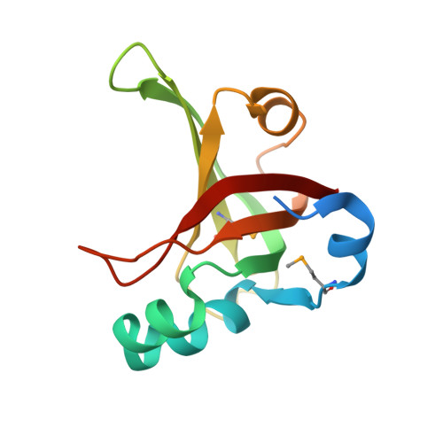
The structure of KPN03535 (gi|152972051), a novel putative lipoprotein from Klebsiella pneumoniae, reveals an OB-fold.
Das, D., Kozbial, P., Han, G.W., Carlton, D., Jaroszewski, L., Abdubek, P., Astakhova, T., Axelrod, H.L., Bakolitsa, C., Chen, C., Chiu, H.J., Chiu, M., Clayton, T., Deller, M.C., Duan, L., Ellrott, K., Elsliger, M.A., Ernst, D., Farr, C.L., Feuerhelm, J., Grzechnik, A., Grant, J.C., Jin, K.K., Johnson, H.A., Klock, H.E., Knuth, M.W., Krishna, S.S., Kumar, A., Marciano, D., McMullan, D., Miller, M.D., Morse, A.T., Nigoghossian, E., Nopakun, A., Okach, L., Oommachen, S., Paulsen, J., Puckett, C., Reyes, R., Rife, C.L., Sefcovic, N., Tien, H.J., Trame, C.B., van den Bedem, H., Weekes, D., Wooten, T., Xu, Q., Hodgson, K.O., Wooley, J., Deacon, A.M., Godzik, A., Lesley, S.A., Wilson, I.A.(2010) Acta Crystallogr Sect F Struct Biol Cryst Commun 66: 1254-1260
- PubMed: 20944219
- DOI: https://doi.org/10.1107/S1744309109018168
- Primary Citation of Related Structures:
3F1Z - PubMed Abstract:
KPN03535 (gi|152972051) is a putative lipoprotein of unknown function that is secreted by Klebsiella pneumoniae MGH 78578. The crystal structure reveals that despite a lack of any detectable sequence similarity to known structures, it is a novel variant of the OB-fold and structurally similar to the bacterial Cpx-pathway protein NlpE, single-stranded DNA-binding (SSB) proteins and toxins. K. pneumoniae MGH 78578 forms part of the normal human skin, mouth and gut flora and is an opportunistic pathogen that is linked to about 8% of all hospital-acquired infections in the USA. This structure provides the foundation for further investigations into this divergent member of the OB-fold family.
Organizational Affiliation:
Stanford Synchrotron Radiation Lightsource, SLAC National Accelerator Laboratory, Menlo Park,California, USA.