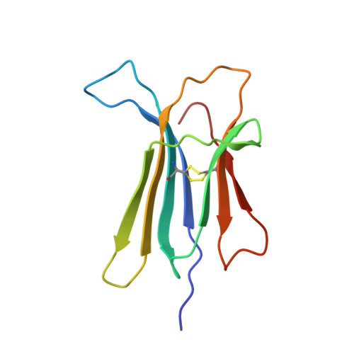
Human beta-2 microglobulin W60V mutant structure: Implications for stability and amyloid aggregation
Ricagno, S., Raimondi, S., Giorgetti, S., Bellotti, V., Bolognesi, M.(2009) Biochem Biophys Res Commun 380: 543-547
- PubMed: 19284997
- DOI: https://doi.org/10.1016/j.bbrc.2009.01.116
- Primary Citation of Related Structures:
3EKC - PubMed Abstract:
Beta-2 microglobulin (?2m) is the light chain of class I major histocompatibility complex (MHC-I). Beta2m is an intrinsically amyloidogenic protein that can assemble into amyloid fibrils in a concentration dependent manner. Beta2m is accumulated in serum of haemodialysed patients, and deposited in the skeletal joints, causing dialysis related amyloidosis. Recent reports suggested that the loop comprised between beta2m strands D and E is crucial for protein stability and for beta2m propensity to aggregate as cross-beta structured fibrils. In particular, the role of Trp60 for beta2m stability has been highlighted by showing that the Trp60-->Gly beta2m mutant is more thermo-stable and less prone to aggregation than the wild type protein. On the contrary the Asp59-->Pro beta2m mutant shows lower Tm and stronger tendency to fibril aggregation. To further analyse such properties, the Trp60-->Val beta2m mutant has been expressed and purified; the propensity to fibrillar aggregation and the folding stability have been assessed, and the X-ray crystal structure determined to 1.8A resolution. The W60V mutant structural features are discussed, focusing on the roles of the DE loop and of residue 60 in relation to ?2m structure and its amyloid aggregation trends.
Organizational Affiliation:
Department of Biomolecular Sciences and Biotechnology, CNR-INFM and CIMAINA, University of Milano, Via Celoria 26, 20133-Milano, Italy.