Structural basis for the secretion of EvpC: a key type VI secretion system protein from Edwardsiella tarda
Jobichen, C., Chakraborty, S., Li, M., Zheng, J., Joseph, L., Mok, Y.K., Leung, K.Y., Sivaraman, J.(2010) PLoS One 5: e12910-e12910
- PubMed: 20886112
- DOI: https://doi.org/10.1371/journal.pone.0012910
- Primary Citation of Related Structures:
3EAA - PubMed Abstract:
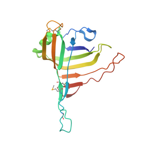
The recently identified type VI secretion system (T6SS) is implicated in the virulence of many gram-negative bacteria. Edwardsiella tarda is an important cause of hemorrhagic septicemia in fish and also gastro- and extra-intestinal infections in humans. The E. tardavirulent protein (EVP) gene cluster encodes a conserved T6SS which contains 16 open reading frames. EvpC is one of the three major EVP secreted proteins and shares high sequence similarity with Hcp1, a key T6SS virulence factor from Pseudomonas aeruginosa. EvpC contributes to the virulence of E. tarda by playing an essential role in functional T6SS. Here, we report the crystal structure of EvpC from E. tarda PPD130/91 at a 2.8 Å resolution, along with functional studies of the protein. EvpC has a β-barrel domain with extended loops. The β-barrel consists of 11 anti-parallel β-strands with an α-helix located on one side. In solution, EvpC exists as a dimer at low concentration and as a hexamer at higher concentration. In the crystal, the symmetry related EvpC molecules form hexameric rings which stack together to form a tube similar to Hcp1. Structure based mutagenesis revealed that N-terminal negatively charged residues, Asp4, Glu15 and Glu26, and C-terminal positively charged residues, Lys161, Lys162 and Lys163, played crucial roles in the secretion of EvpC. Moreover, the localization study indicates the presence of wild type EvpC in cytoplasm, periplasm and secreted fractions, whereas the N-terminal and C-terminal mutants were found mostly in the periplasmic region and was completely absent in the secreted fraction. Results reported here provide insight into the structure, assembly and function of EvpC. Further, these findings can be extended to other EvpC homologs for understanding the mechanism of T6SS and targeting T6SS mediated virulence in gram-negative pathogens.
Organizational Affiliation:
Department of Biological Sciences, National University of Singapore, Singapore, Singapore.