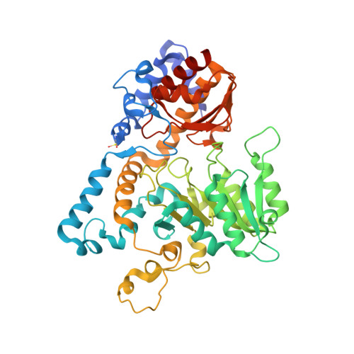
3E9K

Crystal structure of Homo sapiens kynureninase-3-hydroxyhippuric acid inhibitor complex


Experimental Data Snapshot

  • Method: X-RAY DIFFRACTION
  • Resolution: 1.70 Å
  • R-Value Free: 0.191 
  • R-Value Work: 0.153 
  • R-Value Observed: 0.155 

wwPDB Validation   3D Report Full Report


Ligand Structure Quality Assessment 


This is version 1.2 of the entry. See complete history


Literature

Crystal structure of the Homo sapiens kynureninase-3-hydroxyhippuric acid inhibitor complex: insights into the molecular basis of kynureninase substrate specificity.

Lima, S.Kumar, S.Gawandi, V.Momany, C.Phillips, R.S.

(2009) J Med Chem 52: 389-396

  • DOI: https://doi.org/10.1021/jm8010806
  • Primary Citation of Related Structures:  
    3E9K

  • PubMed Abstract: 

    Homo sapiens kynureninase is a pyridoxal-5'-phosphate dependent enzyme that catalyzes the hydrolytic cleavage of 3-hydroxykynurenine to yield 3-hydroxyanthranilate and L-alanine as part of the tryptophan catabolic pathway leading to the de novo biosynthesis of NAD(+). This pathway results in quinolinate, an excitotoxin that is an NMDA receptor agonist. High levels of quinolinate have been correlated with the etiology of neurodegenerative disorders such as AIDS-related dementia and Alzheimer's disease. We have synthesized a novel kynureninase inhibitor, 3-hydroxyhippurate, cocrystallized it with human kynureninase, and solved the atomic structure. On the basis of an analysis of the complex, we designed a series of His-102, Ser-332, and Asn-333 mutants. The H102W/N333T and H102W/S332G/N333T mutants showed complete reversal of substrate specificity between 3-hydroxykynurenine and L-kynurenine, thus defining the primary residues contributing to substrate specificity in kynureninases.


  • Organizational Affiliation

    Department of Biochemistry and Molecular Biology, University of Georgia, Athens, Georgia 30602, USA.


Macromolecules
Find similar proteins by:  (by identity cutoff)  |  3D Structure
Entity ID: 1
MoleculeChains Sequence LengthOrganismDetailsImage
Kynureninase465Homo sapiensMutation(s): 0 
Gene Names: KYNU
EC: 3.7.1.3
UniProt & NIH Common Fund Data Resources
Find proteins for Q16719 (Homo sapiens)
Explore Q16719 
Go to UniProtKB:  Q16719
PHAROS:  Q16719
GTEx:  ENSG00000115919 
Entity Groups  
Sequence Clusters30% Identity50% Identity70% Identity90% Identity95% Identity100% Identity
UniProt GroupQ16719
Sequence Annotations
Expand
  • Reference Sequence
Small Molecules
Ligands 2 Unique
IDChains Name / Formula / InChI Key2D Diagram3D Interactions
PLP
Query on PLP

Download Ideal Coordinates CCD File 
B [auth A]PYRIDOXAL-5'-PHOSPHATE
C8 H10 N O6 P
NGVDGCNFYWLIFO-UHFFFAOYSA-N
3XH
Query on 3XH

Download Ideal Coordinates CCD File 
C [auth A]3-Hydroxyhippuric acid
C9 H9 N O4
XDOFWFNMYJRHEW-UHFFFAOYSA-N
Modified Residues  1 Unique
IDChains TypeFormula2D DiagramParent
CSX
Query on CSX
A
L-PEPTIDE LINKINGC3 H7 N O3 SCYS
Binding Affinity Annotations 
IDSourceBinding Affinity
3XH Binding MOAD:  3E9K Ki: 6.00e+4 (nM) from 1 assay(s)
BindingDB:  3E9K Ki: 6.00e+4 (nM) from 1 assay(s)
Experimental Data & Validation

Experimental Data

  • Method: X-RAY DIFFRACTION
  • Resolution: 1.70 Å
  • R-Value Free: 0.191 
  • R-Value Work: 0.153 
  • R-Value Observed: 0.155 
  • Space Group: C 1 2 1
Unit Cell:
Length ( Å )Angle ( ˚ )
a = 74.444α = 90
b = 77.124β = 109.35
c = 94.558γ = 90
Software Package:
Software NamePurpose
REFMACrefinement
HKL-3000data collection
HKL-3000data reduction
HKL-3000data scaling
PHASERphasing

Structure Validation

View Full Validation Report


Ligand Structure Quality Assessment 


Entry History 

Deposition Data

Revision History  (Full details and data files)

  • Version 1.0: 2008-12-09
    Type: Initial release
  • Version 1.1: 2011-07-13
    Changes: Advisory, Version format compliance
  • Version 1.2: 2023-08-30
    Changes: Data collection, Database references, Derived calculations, Refinement description