3E5V

Crystal Structure Analysis of eqFP611 Double Mutant T122R, N143S


Experimental Data Snapshot

  • Method: X-RAY DIFFRACTION
  • Resolution: 2.10 Å
  • R-Value Free: 0.278 
  • R-Value Work: 0.226 
  • R-Value Observed: 0.228 

wwPDB Validation   3D Report Full Report


This is version 1.6 of the entry. See complete history


Literature

Trans-cis isomerization is responsible for the red-shifted fluorescence in variants of the red fluorescent protein eqFP611.

Nienhaus, K.Nar, H.Heilker, R.Wiedenmann, J.Nienhaus, G.U.

(2008) J Am Chem Soc 130: 12578-12579

  • DOI: https://doi.org/10.1021/ja8046443
  • Primary Citation of Related Structures:  
    3E5T, 3E5V, 3E5W

  • PubMed Abstract: 

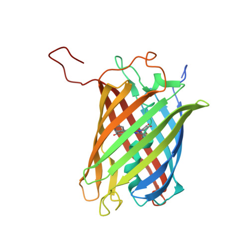
    An important class of red fluorescent proteins (RFPs) feature a 2-iminomethyl-5-(4-hydroxybenzylidene)imidazolinone chromophore. Among these proteins, eqFP611 has the chromophore in a coplanar trans orientation, whereas the cis isomer is preferred by other RFPs such as DsRed and its variants. In the photoactivatable protein asFP595, the chromophore can even be switched from the nonfluorescent trans to the fluorescent cis state by light. By using X-ray crystallography, we have determined the structure of dimeric eqFP611 at high resolution (up to 1.1 A). In the far-red emitting eqFP611 variant d2RFP630, which carries an additional Asn143Ser mutation, the chromophore resides predominantly (approximately 80%) in the cis isomeric state, and in RFP639, which has Asn143Ser and Ser158Cys mutations, the chromophore is found completely in the cis form. The pronounced red shift of excitation and emission maxima of RFP639 can thus unambiguously be assigned to trans-cis isomerization of the chromophore. Among RFPs, eqFP611 is thus unique because its chromophore is highly fluorescent in both the cis and trans isomeric forms.


  • Organizational Affiliation

    Institute of Biophysics, University of Ulm, Albert-Einstein-Allee 11, 89081 Ulm, Germany.


Macromolecules
Find similar proteins by:  (by identity cutoff)  |  3D Structure
Entity ID: 1
MoleculeChains Sequence LengthOrganismDetailsImage
Red fluorescent protein eqFP611242Entacmaea quadricolorMutation(s): 3 
UniProt
Find proteins for Q8ISF8 (Entacmaea quadricolor)
Explore Q8ISF8 
Go to UniProtKB:  Q8ISF8
Entity Groups  
Sequence Clusters30% Identity50% Identity70% Identity90% Identity95% Identity100% Identity
UniProt GroupQ8ISF8
Sequence Annotations
Expand
  • Reference Sequence
Small Molecules
Modified Residues  1 Unique
IDChains TypeFormula2D DiagramParent
NRQ
Query on NRQ
A
L-PEPTIDE LINKINGC16 H17 N3 O4 SMET, TYR, GLY
Experimental Data & Validation

Experimental Data

  • Method: X-RAY DIFFRACTION
  • Resolution: 2.10 Å
  • R-Value Free: 0.278 
  • R-Value Work: 0.226 
  • R-Value Observed: 0.228 
  • Space Group: P 65 2 2
Unit Cell:
Length ( Å )Angle ( ˚ )
a = 62.536α = 90
b = 62.536β = 90
c = 208.022γ = 120
Software Package:
Software NamePurpose
DENZOdata reduction
SCALEPACKdata scaling
TNTrefinement
PDB_EXTRACTdata extraction
BUSTER-TNTrefinement

Structure Validation

View Full Validation Report


Entry History 

Deposition Data

Revision History  (Full details and data files)

  • Version 1.0: 2008-09-23
    Type: Initial release
  • Version 1.1: 2011-07-13
    Changes: Version format compliance
  • Version 1.2: 2011-11-23
    Changes: Structure summary
  • Version 1.3: 2017-10-25
    Changes: Refinement description
  • Version 1.4: 2021-10-20
    Changes: Database references, Derived calculations
  • Version 1.5: 2023-08-30
    Changes: Data collection, Refinement description
  • Version 1.6: 2023-11-15
    Changes: Data collection