3E3C

Structure of GrlR-lipid complex


Experimental Data Snapshot

  • Method: X-RAY DIFFRACTION
  • Resolution: 2.50 Å
  • R-Value Free: 0.278 
  • R-Value Work: 0.231 
  • R-Value Observed: 0.231 

wwPDB Validation   3D Report Full Report


Ligand Structure Quality Assessment 


This is version 1.2 of the entry. See complete history


Literature

Identification and characterization of the lipid binding property of GrlR, a locus of enterocyte effacement regulator.

Jobichen, C.Fernandis, A.Z.Velazquez-Campoy, A.Leung, K.Y.Mok, Y.K.Wenk, M.R.Sivaraman, J.

(2009) Biochem J 

  • DOI: https://doi.org/10.1042/BJ20081588
  • Primary Citation of Related Structures:  
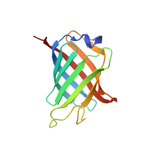
    3E3C

  • PubMed Abstract: 

    Lipocalins are a broad family of proteins identified initially in eukaryotes and more recently in Gram-negative bacteria. The functions of lipocalin or lipid-binding proteins are often elusive and very diverse. Recently, we have determined the structure of GrlR (global regulator of LEE repressor), which plays a key role in the regulation of LEE (locus of enterocyte effacement) proteins. GrlR adopts a lipocalin-like fold that is composed of an eight-stranded beta-barrel followed by an alpha-helix at the C-terminus. GrlR has a highly hydrophobic cavity region and could be a potential transporter of lipophilic molecules. To verify this hypothesis, we carried out structure-based analysis of GrlR, determined the structure of the lipid-GrlR complex and measured the binding of lipid to recombinant GrlR by ITC (isothermal titration calorimetry). In addition, we identified phosphatidylglycerol and phosphatidylethanolamine as the endogenously bound lipid species of GrlR using electrospray-ionization MS. Furthermore, we have shown that the lipid-binding property of GrlR is similar to that of its closest lipocalin structural homologue, beta-lactoglobulin. Our studies demonstrate the hitherto unknown lipid-binding property of GrlR.


  • Organizational Affiliation

    Department of Biological Sciences, National University of Singapore, Singapore.


Macromolecules
Find similar proteins by:  (by identity cutoff)  |  3D Structure
Entity ID: 1
MoleculeChains Sequence LengthOrganismDetailsImage
L0044
A, B
118Escherichia coliMutation(s): 0 
Gene Names: ECs4578
UniProt
Find proteins for Q7DB61 (Escherichia coli O157:H7)
Explore Q7DB61 
Go to UniProtKB:  Q7DB61
Entity Groups  
Sequence Clusters30% Identity50% Identity70% Identity90% Identity95% Identity100% Identity
UniProt GroupQ7DB61
Sequence Annotations
Expand
  • Reference Sequence
Small Molecules
Ligands 1 Unique
IDChains Name / Formula / InChI Key2D Diagram3D Interactions
HHG
Query on HHG

Download Ideal Coordinates CCD File 
C [auth A],
D [auth B]
(2R)-2-HYDROXY-3-(PHOSPHONOOXY)PROPYL HEXANOATE
C9 H19 O7 P
FIMVTNBZKNVWDN-QMMMGPOBSA-N
Binding Affinity Annotations 
IDSourceBinding Affinity
HHG PDBBind:  3E3C Kd: 2400 (nM) from 1 assay(s)
Experimental Data & Validation

Experimental Data

  • Method: X-RAY DIFFRACTION
  • Resolution: 2.50 Å
  • R-Value Free: 0.278 
  • R-Value Work: 0.231 
  • R-Value Observed: 0.231 
  • Space Group: P 21 21 21
Unit Cell:
Length ( Å )Angle ( ˚ )
a = 43.729α = 90
b = 66.015β = 90
c = 83.462γ = 90
Software Package:
Software NamePurpose
ADSCdata collection
PHASERphasing
CNSrefinement
HKL-2000data reduction
HKL-2000data scaling

Structure Validation

View Full Validation Report


Ligand Structure Quality Assessment 


Entry History 

Deposition Data

Revision History  (Full details and data files)

  • Version 1.0: 2009-04-07
    Type: Initial release
  • Version 1.1: 2011-07-13
    Changes: Version format compliance
  • Version 1.2: 2023-11-01
    Changes: Data collection, Database references, Derived calculations, Refinement description