3E20

Crystal structure of S.pombe eRF1/eRF3 complex


Experimental Data Snapshot

  • Method: X-RAY DIFFRACTION
  • Resolution: 3.50 Å
  • R-Value Free: 0.280 
  • R-Value Work: 0.258 
  • R-Value Observed: 0.259 

wwPDB Validation   3D Report Full Report


This is version 1.2 of the entry. See complete history


Literature

Structural insights into eRF3 and stop codon recognition by eRF1

Cheng, Z.Saito, K.Pisarev, A.V.Wada, M.Pisareva, V.P.Pestova, T.V.Gajda, M.Round, A.Kong, C.Lim, M.Nakamura, Y.Svergun, D.I.Ito, K.Song, H.

(2009) Genes Dev 23: 1106-1118

  • DOI: https://doi.org/10.1101/gad.1770109
  • Primary Citation of Related Structures:  
    3E1Y, 3E20

  • PubMed Abstract: 

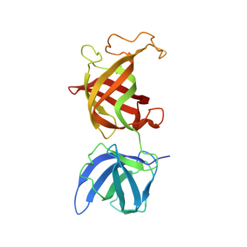
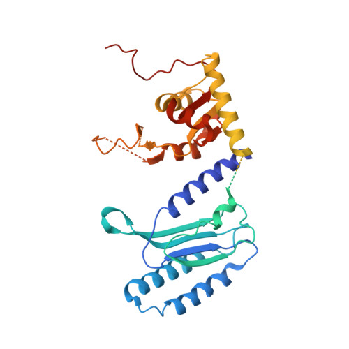
    Eukaryotic translation termination is mediated by two interacting release factors, eRF1 and eRF3, which act cooperatively to ensure efficient stop codon recognition and fast polypeptide release. The crystal structures of human and Schizosaccharomyces pombe full-length eRF1 in complex with eRF3 lacking the GTPase domain revealed details of the interaction between these two factors and marked conformational changes in eRF1 that occur upon binding to eRF3, leading eRF1 to resemble a tRNA molecule. Small-angle X-ray scattering analysis of the eRF1/eRF3/GTP complex suggested that eRF1's M domain contacts eRF3's GTPase domain. Consistently, mutation of Arg192, which is predicted to come in close contact with the switch regions of eRF3, revealed its important role for eRF1's stimulatory effect on eRF3's GTPase activity. An ATP molecule used as a crystallization additive was bound in eRF1's putative decoding area. Mutational analysis of the ATP-binding site shed light on the mechanism of stop codon recognition by eRF1.


  • Organizational Affiliation

    Cancer and Developmental Cell Biology Division, Institute of Molecular and Cell Biology, Agency for Science, Technology, and Research (A*STAR), Singapore, Singapore.


Macromolecules
Find similar proteins by:  (by identity cutoff)  |  3D Structure
Entity ID: 1
MoleculeChains Sequence LengthOrganismDetailsImage
Eukaryotic peptide chain release factor GTP-binding subunitA,
C [auth D],
E,
G [auth J]
201Schizosaccharomyces pombeMutation(s): 0 
Gene Names: sup35SPCC584.04
UniProt
Find proteins for O74718 (Schizosaccharomyces pombe (strain 972 / ATCC 24843))
Explore O74718 
Go to UniProtKB:  O74718
Entity Groups  
Sequence Clusters30% Identity50% Identity70% Identity90% Identity95% Identity100% Identity
UniProt GroupO74718
Sequence Annotations
Expand
  • Reference Sequence
Find similar proteins by:  (by identity cutoff)  |  3D Structure
Entity ID: 2
MoleculeChains Sequence LengthOrganismDetailsImage
Eukaryotic peptide chain release factor subunit 1B [auth C],
D [auth B],
F [auth H],
H [auth K]
441Schizosaccharomyces pombeMutation(s): 0 
Gene Names: sup45SPAC1834.01
UniProt
Find proteins for P79063 (Schizosaccharomyces pombe (strain 972 / ATCC 24843))
Explore P79063 
Go to UniProtKB:  P79063
Entity Groups  
Sequence Clusters30% Identity50% Identity70% Identity90% Identity95% Identity100% Identity
UniProt GroupP79063
Sequence Annotations
Expand
  • Reference Sequence
Experimental Data & Validation

Experimental Data

  • Method: X-RAY DIFFRACTION
  • Resolution: 3.50 Å
  • R-Value Free: 0.280 
  • R-Value Work: 0.258 
  • R-Value Observed: 0.259 
  • Space Group: P 43
Unit Cell:
Length ( Å )Angle ( ˚ )
a = 129.848α = 90
b = 129.848β = 90
c = 332.638γ = 90
Software Package:
Software NamePurpose
REFMACrefinement
ADSCdata collection
MOSFLMdata reduction
SCALAdata scaling
PHASERphasing

Structure Validation

View Full Validation Report


Entry History 

Deposition Data

Revision History  (Full details and data files)

  • Version 1.0: 2009-05-19
    Type: Initial release
  • Version 1.1: 2011-07-13
    Changes: Advisory, Version format compliance
  • Version 1.2: 2023-11-01
    Changes: Data collection, Database references, Refinement description