3DVG

Crystal structure of K63-specific fab Apu.3A8 bound to K63-linked di-ubiquitin


Experimental Data Snapshot

  • Method: X-RAY DIFFRACTION
  • Resolution: 2.60 Å
  • R-Value Free: 0.261 
  • R-Value Work: 0.219 
  • R-Value Observed: 0.224 

wwPDB Validation   3D Report Full Report


This is version 1.2 of the entry. See complete history


Literature

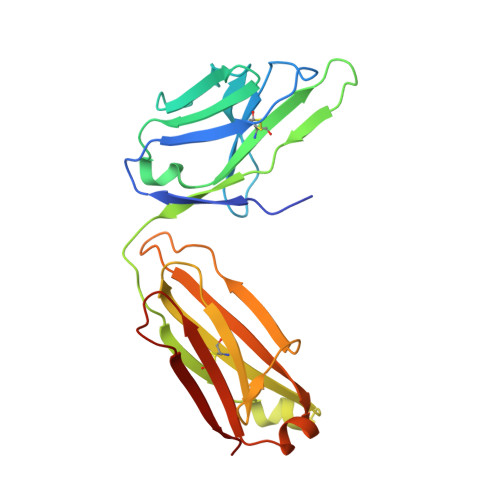
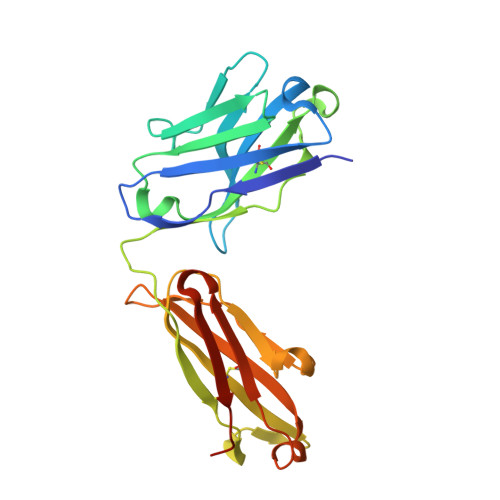
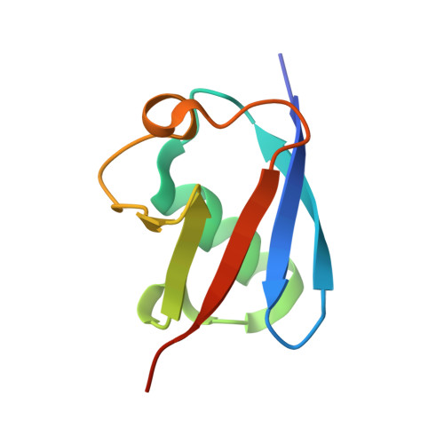
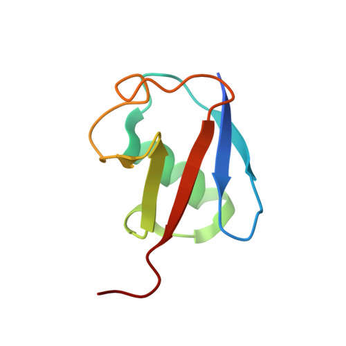
Ubiquitin chain editing revealed by polyubiquitin linkage-specific antibodies.

Newton, K.Matsumoto, M.L.Wertz, I.E.Kirkpatrick, D.S.Lill, J.R.Tan, J.Dugger, D.Gordon, N.Sidhu, S.S.Fellouse, F.A.Komuves, L.French, D.M.Ferrando, R.E.Lam, C.Compaan, D.Yu, C.Bosanac, I.Hymowitz, S.G.Kelley, R.F.Dixit, V.M.

(2008) Cell 134: 668-678

  • DOI: https://doi.org/10.1016/j.cell.2008.07.039
  • Primary Citation of Related Structures:  
    3DVG, 3DVN

  • PubMed Abstract: 

    Posttranslational modification of proteins with polyubiquitin occurs in diverse signaling pathways and is tightly regulated to ensure cellular homeostasis. Studies employing ubiquitin mutants suggest that the fate of polyubiquitinated proteins is determined by which lysine within ubiquitin is linked to the C terminus of an adjacent ubiquitin. We have developed linkage-specific antibodies that recognize polyubiquitin chains joined through lysine 63 (K63) or 48 (K48). A cocrystal structure of an anti-K63 linkage Fab bound to K63-linked diubiquitin provides insight into the molecular basis for specificity. We use these antibodies to demonstrate that RIP1, which is essential for tumor necrosis factor-induced NF-kappaB activation, and IRAK1, which participates in signaling by interleukin-1beta and Toll-like receptors, both undergo polyubiquitin editing in stimulated cells. Both kinase adaptors initially acquire K63-linked polyubiquitin, while at later times K48-linked polyubiquitin targets them for proteasomal degradation. Polyubiquitin editing may therefore be a general mechanism for attenuating innate immune signaling.


  • Organizational Affiliation

    Department of Physiological Chemistry, Genentech, Inc., 1 DNA Way, South San Francisco, CA 94080, USA.


Macromolecules
Find similar proteins by:  (by identity cutoff)  |  3D Structure
Entity ID: 1
MoleculeChains Sequence LengthOrganismDetailsImage
Human IgG1 fab fragment light chain217Homo sapiensMutation(s): 0 
Gene Names: fab fragment light chain
UniProt
Find proteins for Q6PIL8 (Homo sapiens)
Explore Q6PIL8 
Go to UniProtKB:  Q6PIL8
Entity Groups  
Sequence Clusters30% Identity50% Identity70% Identity90% Identity95% Identity100% Identity
UniProt GroupQ6PIL8
Sequence Annotations
Expand
  • Reference Sequence
Find similar proteins by:  (by identity cutoff)  |  3D Structure
Entity ID: 2
MoleculeChains Sequence LengthOrganismDetailsImage
Human IgG1 fab fragment heavy chain230Homo sapiensMutation(s): 0 
Gene Names: fab fragment light chain
Entity Groups  
Sequence Clusters30% Identity50% Identity70% Identity90% Identity95% Identity100% Identity
Sequence Annotations
Expand
  • Reference Sequence
Find similar proteins by:  (by identity cutoff)  |  3D Structure
Entity ID: 3
MoleculeChains Sequence LengthOrganismDetailsImage
Ubiquitin D77C [auth X]80Homo sapiensMutation(s): 1 
Gene Names: RPS27AUBA80UBCEP1UBA52UBCEP2UBBUBC
UniProt & NIH Common Fund Data Resources
Find proteins for P0CG48 (Homo sapiens)
Explore P0CG48 
Go to UniProtKB:  P0CG48
PHAROS:  P0CG48
GTEx:  ENSG00000150991 
Entity Groups  
Sequence Clusters30% Identity50% Identity70% Identity90% Identity95% Identity100% Identity
UniProt GroupP0CG48
Sequence Annotations
Expand
  • Reference Sequence
Find similar proteins by:  (by identity cutoff)  |  3D Structure
Entity ID: 4
MoleculeChains Sequence LengthOrganismDetailsImage
UbiquitinD [auth Y]79Homo sapiensMutation(s): 1 
Gene Names: RPS27AUBA80UBCEP1UBA52UBCEP2UBBUBC
UniProt & NIH Common Fund Data Resources
Find proteins for P0CG48 (Homo sapiens)
Explore P0CG48 
Go to UniProtKB:  P0CG48
PHAROS:  P0CG48
GTEx:  ENSG00000150991 
Entity Groups  
Sequence Clusters30% Identity50% Identity70% Identity90% Identity95% Identity100% Identity
UniProt GroupP0CG48
Sequence Annotations
Expand
  • Reference Sequence
Experimental Data & Validation

Experimental Data

  • Method: X-RAY DIFFRACTION
  • Resolution: 2.60 Å
  • R-Value Free: 0.261 
  • R-Value Work: 0.219 
  • R-Value Observed: 0.224 
  • Space Group: C 1 2 1
Unit Cell:
Length ( Å )Angle ( ˚ )
a = 106.806α = 90
b = 88.117β = 108.28
c = 90.226γ = 90
Software Package:
Software NamePurpose
REFMACrefinement
Blu-Icedata collection
HKL-2000data reduction
HKL-2000data scaling
PHASERphasing

Structure Validation

View Full Validation Report


Entry History 

Deposition Data

Revision History  (Full details and data files)

  • Version 1.0: 2008-09-30
    Type: Initial release
  • Version 1.1: 2011-07-13
    Changes: Advisory, Refinement description, Version format compliance
  • Version 1.2: 2021-10-20
    Changes: Database references, Derived calculations