3DSF

Crystal structure of anti-osteopontin antibody 23C3 in complex with W43A mutated epitope peptide


Experimental Data Snapshot

  • Method: X-RAY DIFFRACTION
  • Resolution: 2.80 Å
  • R-Value Free: 0.297 
  • R-Value Work: 0.241 
  • R-Value Observed: 0.244 

wwPDB Validation   3D Report Full Report


This is version 2.2 of the entry. See complete history


Literature

Molecular basis of recognition of human osteopontin by 23C3, a potential therapeutic antibody for treatment of rheumatoid arthritis

Du, J.Hou, S.Zhong, C.Lai, Z.Yang, H.Dai, J.Zhang, D.Wang, H.Guo, Y.Ding, J.

(2008) J Mol Biol 382: 835-842

  • DOI: https://doi.org/10.1016/j.jmb.2008.07.075
  • Primary Citation of Related Structures:  
    3CXD, 3DSF

  • PubMed Abstract: 

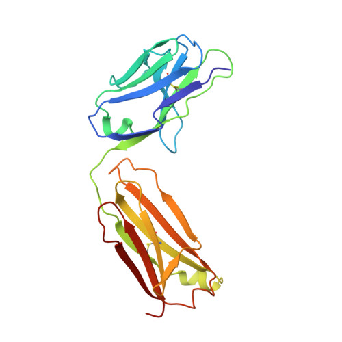
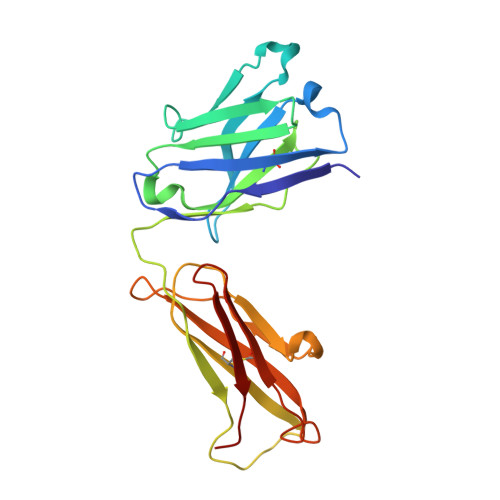
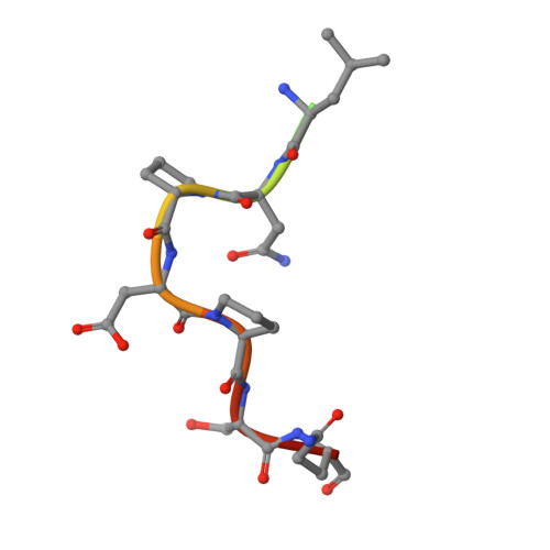
    Osteopontin plays an important role in the development and perpetuation of rheumatoid arthritis (RA). Antibodies targeting osteopontin have shown promising therapeutic benefits against this disease. We have previously reported a novel anti-RA monoclonal antibody, namely, 23C3, and shown it capable of alleviating the symptoms of RA in a murine collagen-induced arthritis model, restoring the cytokine production profile in joint tissues, and reducing T-cell recall responses to collagen type II. We describe here the crystal structure of 23C3 in complex with its epitope peptide. Analyses of the complex structure reveal the molecular mechanism of osteopontin recognition by 23C3. The peptide folds into two tandem beta-turns, and two key residues of the peptide are identified to be critical for the recognition by 23C3: TrpP43 is deeply embedded into a hydrophobic pocket formed by AlaL34, TyrL36, LeuL46, TyrL49, PheL91, and MetH102 and therefore has extensive hydrophobic interactions with 23C3, while AspP47 has a network of hydrophilic interactions with residues ArgH50, ArgH52, SerH53, and AsnH56 of the antibody. Besides the complementarity-determining region loops, the framework region L2 of 23C3 is also shown to interact with the epitope peptide, which is not common in the antibody-antigen interactions and thus could be exploited in the engineering of 23C3. These results not only provide valuable information for further improvement of 23C3 such as chimerization or humanization for its therapeutic application, but also reveal the features of this specific epitope of osteopontin that may be useful for the development of new antibody drugs against RA.


  • Organizational Affiliation

    State Key Laboratory of Molecular Biology, Institute of Biochemistry and Cell Biology, Shanghai Institutes for Biological Sciences, Chinese Academy of Sciences, 320 Yue-Yang Road, Shanghai 200031, China.


Macromolecules
Find similar proteins by:  (by identity cutoff)  |  3D Structure
Entity ID: 1
MoleculeChains Sequence LengthOrganismDetailsImage
Fab fragment of anti-osteopontin antibody 23C3, Light chainA [auth L]213Mus musculusMutation(s): 0 
Entity Groups  
Sequence Clusters30% Identity50% Identity70% Identity90% Identity95% Identity100% Identity
Sequence Annotations
Expand
  • Reference Sequence
Find similar proteins by:  (by identity cutoff)  |  3D Structure
Entity ID: 2
MoleculeChains Sequence LengthOrganismDetailsImage
Fab fragment of anti-osteopontin antibody 23C3, Heavy chainB [auth H]216Mus musculusMutation(s): 0 
Entity Groups  
Sequence Clusters30% Identity50% Identity70% Identity90% Identity95% Identity100% Identity
Sequence Annotations
Expand
  • Reference Sequence

Find similar proteins by:  Sequence   |   3D Structure  

Entity ID: 3
MoleculeChains Sequence LengthOrganismDetailsImage
a peptide from osteopontinC [auth P]12N/AMutation(s): 1 
UniProt & NIH Common Fund Data Resources
Find proteins for P10451 (Homo sapiens)
Explore P10451 
Go to UniProtKB:  P10451
PHAROS:  P10451
GTEx:  ENSG00000118785 
Entity Groups  
Sequence Clusters30% Identity50% Identity70% Identity90% Identity95% Identity100% Identity
UniProt GroupP10451
Sequence Annotations
Expand
  • Reference Sequence
Oligosaccharides

Help

Entity ID: 4
MoleculeChains Length2D Diagram Glycosylation3D Interactions
alpha-D-mannopyranose-(1-3)-beta-D-mannopyranose-(1-4)-2-acetamido-2-deoxy-beta-D-glucopyranose-(1-4)-2-acetamido-2-deoxy-beta-D-glucopyranoseD [auth A]4N-Glycosylation
Glycosylation Resources
GlyTouCan:  G81315DD
GlyCosmos:  G81315DD
GlyGen:  G81315DD
Experimental Data & Validation

Experimental Data

  • Method: X-RAY DIFFRACTION
  • Resolution: 2.80 Å
  • R-Value Free: 0.297 
  • R-Value Work: 0.241 
  • R-Value Observed: 0.244 
  • Space Group: P 21 21 21
Unit Cell:
Length ( Å )Angle ( ˚ )
a = 41.73α = 90
b = 92.058β = 90
c = 122.104γ = 90
Software Package:
Software NamePurpose
REFMACrefinement
CrystalCleardata collection
CrystalCleardata reduction
CrystalCleardata scaling
PHASERphasing

Structure Validation

View Full Validation Report


Entry History 

Deposition Data

Revision History  (Full details and data files)

  • Version 1.0: 2008-10-14
    Type: Initial release
  • Version 1.1: 2011-07-13
    Changes: Non-polymer description, Version format compliance
  • Version 2.0: 2020-07-29
    Type: Remediation
    Reason: Carbohydrate remediation
    Changes: Atomic model, Data collection, Derived calculations, Structure summary
  • Version 2.1: 2021-11-10
    Changes: Database references, Structure summary
  • Version 2.2: 2023-11-01
    Changes: Data collection, Refinement description