3DDC

Crystal Structure of NORE1A in Complex with RAS


Experimental Data Snapshot

  • Method: X-RAY DIFFRACTION
  • Resolution: 1.80 Å
  • R-Value Free: 0.230 
  • R-Value Work: 0.193 
  • R-Value Observed: 0.195 

wwPDB Validation   3D Report Full Report


Ligand Structure Quality Assessment 


This is version 1.3 of the entry. See complete history


Literature

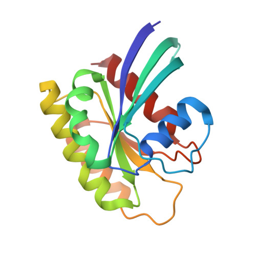
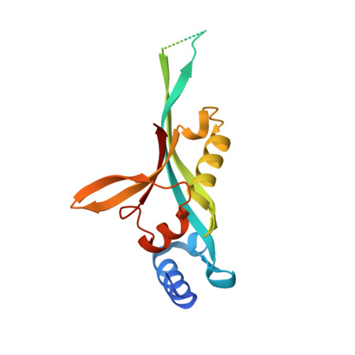
Novel type of Ras effector interaction established between tumour suppressor NORE1A and Ras switch II

Stieglitz, B.Bee, C.Schwarz, D.Yildiz, O.Moshnikova, A.Khokhlatchev, A.Herrmann, C.

(2008) EMBO J 27: 1995-2005

  • DOI: https://doi.org/10.1038/emboj.2008.125
  • Primary Citation of Related Structures:  
    3DDC

  • PubMed Abstract: 

    A class of putative Ras effectors called Ras association domain family (RASSF) represents non-enzymatic adaptors that were shown to be important in tumour suppression. RASSF5, a member of this family, exists in two splice variants known as NORE1A and RAPL. Both of them are involved in distinct cellular pathways triggered by Ras and Rap, respectively. Here we describe the crystal structure of Ras in complex with the Ras binding domain (RBD) of NORE1A/RAPL. All Ras effectors share a common topology in their RBD creating an interface with the switch I region of Ras, whereas NORE1A/RAPL RBD reveals additional structural elements forming a unique Ras switch II binding site. Consequently, the contact area of NORE1A is extended as compared with other Ras effectors. We demonstrate that the enlarged interface provides a rationale for an exceptionally long lifetime of the complex. This is a specific attribute characterizing the effector function of NORE1A/RAPL as adaptors, in contrast to classical enzymatic effectors such as Raf, RalGDS or PI3K, which are known to form highly dynamic short-lived complexes with Ras.


  • Organizational Affiliation

    Physikalische Chemie 1, Fakultät für Chemie und Biochemie, Ruhr-Universität Bochum, Bochum, Germany.


Macromolecules
Find similar proteins by:  (by identity cutoff)  |  3D Structure
Entity ID: 1
MoleculeChains Sequence LengthOrganismDetailsImage
GTPase HRas166Homo sapiensMutation(s): 2 
Gene Names: HRASHRAS1
EC: 3.6.5.2
UniProt & NIH Common Fund Data Resources
Find proteins for P01112 (Homo sapiens)
Explore P01112 
Go to UniProtKB:  P01112
PHAROS:  P01112
GTEx:  ENSG00000174775 
Entity Groups  
Sequence Clusters30% Identity50% Identity70% Identity90% Identity95% Identity100% Identity
UniProt GroupP01112
Sequence Annotations
Expand
  • Reference Sequence
Find similar proteins by:  (by identity cutoff)  |  3D Structure
Entity ID: 2
MoleculeChains Sequence LengthOrganismDetailsImage
Ras association domain-containing family protein 5163Mus musculusMutation(s): 2 
Gene Names: Rassf5Nore1Rapl
UniProt
Find proteins for Q5EBH1 (Mus musculus)
Explore Q5EBH1 
Go to UniProtKB:  Q5EBH1
Entity Groups  
Sequence Clusters30% Identity50% Identity70% Identity90% Identity95% Identity100% Identity
UniProt GroupQ5EBH1
Sequence Annotations
Expand
  • Reference Sequence
Small Molecules
Ligands 2 Unique
IDChains Name / Formula / InChI Key2D Diagram3D Interactions
GNP
Query on GNP

Download Ideal Coordinates CCD File 
D [auth A]PHOSPHOAMINOPHOSPHONIC ACID-GUANYLATE ESTER
C10 H17 N6 O13 P3
UQABYHGXWYXDTK-UUOKFMHZSA-N
MG
Query on MG

Download Ideal Coordinates CCD File 
C [auth A]MAGNESIUM ION
Mg
JLVVSXFLKOJNIY-UHFFFAOYSA-N
Experimental Data & Validation

Experimental Data

  • Method: X-RAY DIFFRACTION
  • Resolution: 1.80 Å
  • R-Value Free: 0.230 
  • R-Value Work: 0.193 
  • R-Value Observed: 0.195 
  • Space Group: C 1 2 1
Unit Cell:
Length ( Å )Angle ( ˚ )
a = 79.6α = 90
b = 88β = 125
c = 56.5γ = 90
Software Package:
Software NamePurpose
AMoREphasing
REFMACrefinement
ADSCdata collection
XDSdata reduction
XSCALEdata scaling

Structure Validation

View Full Validation Report


Ligand Structure Quality Assessment 


Entry History 

Deposition Data

Revision History  (Full details and data files)

  • Version 1.0: 2008-07-15
    Type: Initial release
  • Version 1.1: 2011-07-13
    Changes: Advisory, Version format compliance
  • Version 1.2: 2011-07-20
    Changes: Refinement description
  • Version 1.3: 2023-11-01
    Changes: Data collection, Database references, Derived calculations, Refinement description