3DAJ

Crystal structure of Aurora A complexed with an inhibitor discovered through site-directed dynamic tethering


Experimental Data Snapshot

  • Method: X-RAY DIFFRACTION
  • Resolution: 2.00 Å
  • R-Value Free: 0.284 
  • R-Value Work: 0.251 
  • R-Value Observed: 0.253 

wwPDB Validation   3D Report Full Report


This is version 1.3 of the entry. See complete history


Literature

Discovery of an Aurora kinase inhibitor through site-specific dynamic combinatorial chemistry.

Cancilla, M.T.He, M.M.Viswanathan, N.Simmons, R.L.Taylor, M.Fung, A.D.Cao, K.Erlanson, D.A.

(2008) Bioorg Med Chem Lett 18: 3978-3981

  • DOI: https://doi.org/10.1016/j.bmcl.2008.06.011
  • Primary Citation of Related Structures:  
    3DAJ

  • PubMed Abstract: 

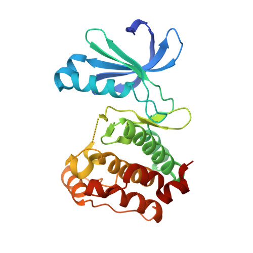
    We demonstrate a fragment-based lead discovery method that combines site-directed ligand discovery with dynamic combinatorial chemistry. Our technique targets dynamic combinatorial screening to a specified region of a protein by using reversible disulfide chemistry. We have used this technology to rapidly identify inhibitors of the drug target Aurora A that span the purine-binding site and the adaptive pocket of the kinase. The binding mode of a noncovalent inhibitor has been further characterized through crystallography.


  • Organizational Affiliation

    Sunesis Pharmaceuticals, Inc., 395 Oyster Point Boulevard, Suite 400, South San Francisco, CA 94080, USA.


Macromolecules
Find similar proteins by:  (by identity cutoff)  |  3D Structure
Entity ID: 1
MoleculeChains Sequence LengthOrganismDetailsImage
serine/threonine kinase 6272Mus musculusMutation(s): 4 
Gene Names: AurkaStk6
UniProt & NIH Common Fund Data Resources
Find proteins for P97477 (Mus musculus)
Explore P97477 
Go to UniProtKB:  P97477
IMPC:  MGI:894678
Entity Groups  
Sequence Clusters30% Identity50% Identity70% Identity90% Identity95% Identity100% Identity
UniProt GroupP97477
Sequence Annotations
Expand
  • Reference Sequence
Small Molecules
Ligands 1 Unique
IDChains Name / Formula / InChI Key2D Diagram3D Interactions
FXG
Query on FXG

Download Ideal Coordinates CCD File 
B [auth A]N-butyl-3-{[6-(9H-purin-6-ylamino)hexanoyl]amino}benzamide
C22 H29 N7 O2
WOYITRCGMUXUDE-UHFFFAOYSA-N
Binding Affinity Annotations 
IDSourceBinding Affinity
FXG Binding MOAD:  3DAJ IC50: 2900 (nM) from 1 assay(s)
PDBBind:  3DAJ IC50: 2900 (nM) from 1 assay(s)
Experimental Data & Validation

Experimental Data

  • Method: X-RAY DIFFRACTION
  • Resolution: 2.00 Å
  • R-Value Free: 0.284 
  • R-Value Work: 0.251 
  • R-Value Observed: 0.253 
  • Space Group: P 41 21 2
Unit Cell:
Length ( Å )Angle ( ˚ )
a = 84.932α = 90
b = 84.932β = 90
c = 77.025γ = 90
Software Package:
Software NamePurpose
REFMACrefinement
MAR345dtbdata collection
MOSFLMdata reduction
SCALAdata scaling
AMoREphasing

Structure Validation

View Full Validation Report


Entry History 

Deposition Data

  • Released Date: 2008-07-08 
  • Deposition Author(s): He, M.M.

Revision History  (Full details and data files)

  • Version 1.0: 2008-07-08
    Type: Initial release
  • Version 1.1: 2011-07-13
    Changes: Version format compliance
  • Version 1.2: 2021-10-20
    Changes: Database references, Derived calculations
  • Version 1.3: 2024-02-21
    Changes: Data collection