3D6I

Structure of the Thioredoxin-like Domain of Yeast Glutaredoxin 3


Experimental Data Snapshot

  • Method: X-RAY DIFFRACTION
  • Resolution: 1.50 Å
  • R-Value Free: 0.249 
  • R-Value Work: 0.223 
  • R-Value Observed: 0.225 

wwPDB Validation   3D Report Full Report


This is version 1.3 of the entry. See complete history


Literature

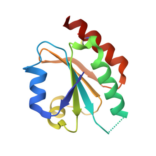
Structure of the thioredoxin-like domain of yeast glutaredoxin 3.

Gibson, L.M.Dingra, N.N.Outten, C.E.Lebioda, L.

(2008) Acta Crystallogr D Biol Crystallogr 64: 927-932

  • DOI: https://doi.org/10.1107/S0907444908021641
  • Primary Citation of Related Structures:  
    3D6I

  • PubMed Abstract: 

    Yeast glutaredoxin 3 (Grx3) is a cytosolic protein that regulates the activity of the iron-responsive transcriptional activator Aft1. This member of the monothiol glutaredoxin family contains a thioredoxin-like domain and a glutaredoxin-like domain, which both possess a monothiol active site. The crystal structure of the thioredoxin-like domain has been determined at 1.5 A resolution and represents the first published structure of this domain for the monothiol glutaredoxin family. The loop containing the signature motif WAxxC is partially disordered, indicating a greater degree of flexibility in this region compared with classical dithiol thioredoxins with a WCGPC active-site motif.


  • Organizational Affiliation

    Department of Chemistry and Biochemistry, University of South Carolina, Columbia, SC 29208, USA.


Macromolecules
Find similar proteins by:  (by identity cutoff)  |  3D Structure
Entity ID: 1
MoleculeChains Sequence LengthOrganismDetailsImage
Monothiol glutaredoxin-3
A, B
112Saccharomyces cerevisiaeMutation(s): 0 
Gene Names: GRX3YDR098CYD8557.05C
UniProt
Find proteins for Q03835 (Saccharomyces cerevisiae (strain ATCC 204508 / S288c))
Explore Q03835 
Go to UniProtKB:  Q03835
Entity Groups  
Sequence Clusters30% Identity50% Identity70% Identity90% Identity95% Identity100% Identity
UniProt GroupQ03835
Sequence Annotations
Expand
  • Reference Sequence
Small Molecules
Modified Residues  1 Unique
IDChains TypeFormula2D DiagramParent
CME
Query on CME
A, B
L-PEPTIDE LINKINGC5 H11 N O3 S2CYS
Experimental Data & Validation

Experimental Data

  • Method: X-RAY DIFFRACTION
  • Resolution: 1.50 Å
  • R-Value Free: 0.249 
  • R-Value Work: 0.223 
  • R-Value Observed: 0.225 
  • Space Group: P 1 21 1
Unit Cell:
Length ( Å )Angle ( ˚ )
a = 47.906α = 90
b = 48.611β = 116.76
c = 55.294γ = 90
Software Package:
Software NamePurpose
DENZOdata reduction
SCALEPACKdata scaling
REFMACrefinement
PDB_EXTRACTdata extraction
SERGUIdata collection
HKL-2000data reduction
AMoREphasing

Structure Validation

View Full Validation Report


Entry History 

Deposition Data

Revision History  (Full details and data files)

  • Version 1.0: 2008-09-02
    Type: Initial release
  • Version 1.1: 2011-07-13
    Changes: Version format compliance
  • Version 1.2: 2017-10-25
    Changes: Refinement description
  • Version 1.3: 2023-08-30
    Changes: Data collection, Database references, Derived calculations, Refinement description