3D0V

Crystal structure of the HIV-1 Cross Neutralizing Monoclonal Antibody 2F5 in complex with gp41 Peptide LLELDKWASLW


Experimental Data Snapshot

  • Method: X-RAY DIFFRACTION
  • Resolution: 2.05 Å
  • R-Value Free: 0.232 
  • R-Value Work: 0.218 
  • R-Value Observed: 0.218 

wwPDB Validation   3D Report Full Report


This is version 1.2 of the entry. See complete history


Literature

Structural details of HIV-1 recognition by the broadly neutralizing monoclonal antibody 2F5: epitope conformation, antigen-recognition loop mobility, and anion-binding site.

Julien, J.P.Bryson, S.Nieva, J.L.Pai, E.F.

(2008) J Mol Biol 384: 377-392

  • DOI: https://doi.org/10.1016/j.jmb.2008.09.024
  • Primary Citation of Related Structures:  
    2P8L, 2P8M, 2P8P, 2PR4, 3D0L, 3D0V, 3DRO, 3DRQ

  • PubMed Abstract: 

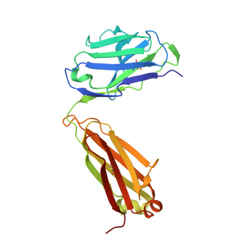
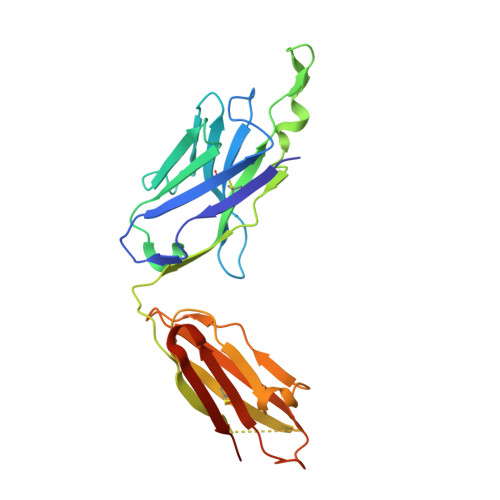
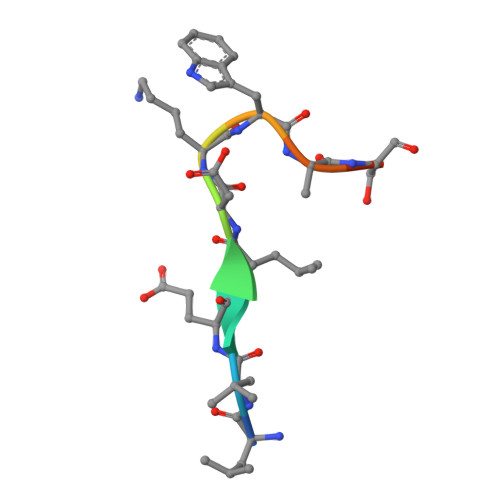
    2F5 is a monoclonal antibody with potent and broadly neutralizing activity against HIV-1. It targets the membrane-proximal external region (MPER) of the gp41 subunit of the envelope glycoprotein and interferes with the process of fusion between viral and host cell membranes. This study presents eight 2F5 F(ab)' crystal structures in complex with various gp41 peptide epitopes. These structures reveal several key features of this antibody-antigen interaction. (1) Whenever free of contacts caused by crystal artifacts, the extended complementarity-determining region H3 loop is mobile; this is true for ligand-free and epitope-bound forms. (2) The interaction between the antibody and the gp41 ELDKWA epitope core is absolutely critical, and there are also close and specific contacts with residues located N-terminal to the epitope core. (3) Residues located at the C-terminus of the gp41 ELDKWA core do not interact as tightly with the antibody. However, in the presence of a larger peptide containing the gp41 fusion peptide segment, these residues adopt a conformation consistent with the start of an alpha-helix. (4) At high sulfate concentrations, the electron density maps of 2F5 F(ab)'-peptide complexes contain a peak that may mark a binding site for phosphate groups of negatively charged lipid headgroups. The refined atomic-level details of 2F5 paratope-epitope interactions revealed here should contribute to a better understanding of the mechanism of 2F5-based virus neutralization, in general, and prove important for the design of potential vaccine candidates intended to elicit 2F5-like antibody production.


  • Organizational Affiliation

    Department of Biochemistry, University of Toronto, 1 King's College Circle, Toronto, Ontario, Canada M5S 1A8.


Macromolecules
Find similar proteins by:  (by identity cutoff)  |  3D Structure
Entity ID: 1
MoleculeChains Sequence LengthOrganismDetailsImage
2F5 Fab heavy chain214Homo sapiensMutation(s): 0 
Entity Groups  
Sequence Clusters30% Identity50% Identity70% Identity90% Identity95% Identity100% Identity
Sequence Annotations
Expand
  • Reference Sequence
Find similar proteins by:  (by identity cutoff)  |  3D Structure
Entity ID: 2
MoleculeChains Sequence LengthOrganismDetailsImage
2F5 Fab light chain237Homo sapiensMutation(s): 0 
UniProt
Find proteins for Q8NF17 (Homo sapiens)
Explore Q8NF17 
Go to UniProtKB:  Q8NF17
Entity Groups  
Sequence Clusters30% Identity50% Identity70% Identity90% Identity95% Identity100% Identity
UniProt GroupQ8NF17
Sequence Annotations
Expand
  • Reference Sequence

Find similar proteins by:  Sequence   |   3D Structure  

Entity ID: 3
MoleculeChains Sequence LengthOrganismDetailsImage
gp41 peptide LLELDKWASLW11N/AMutation(s): 0 
UniProt
Find proteins for P04578 (Human immunodeficiency virus type 1 group M subtype B (isolate HXB2))
Explore P04578 
Go to UniProtKB:  P04578
Entity Groups  
Sequence Clusters30% Identity50% Identity70% Identity90% Identity95% Identity100% Identity
UniProt GroupP04578
Sequence Annotations
Expand
  • Reference Sequence
Experimental Data & Validation

Experimental Data

  • Method: X-RAY DIFFRACTION
  • Resolution: 2.05 Å
  • R-Value Free: 0.232 
  • R-Value Work: 0.218 
  • R-Value Observed: 0.218 
  • Space Group: P 21 21 21
Unit Cell:
Length ( Å )Angle ( ˚ )
a = 58.2α = 90
b = 64.6β = 90
c = 177.7γ = 90
Software Package:
Software NamePurpose
CNSrefinement
DENZOdata reduction
SCALEPACKdata scaling
CNSphasing

Structure Validation

View Full Validation Report


Entry History 

Deposition Data

Revision History  (Full details and data files)

  • Version 1.0: 2008-07-29
    Type: Initial release
  • Version 1.1: 2011-07-13
    Changes: Version format compliance
  • Version 1.2: 2023-08-30
    Changes: Data collection, Database references, Refinement description