3COO

The crystal structure of Reelin-N domain of F-spondin


Experimental Data Snapshot

  • Method: X-RAY DIFFRACTION
  • Resolution: 2.00 Å
  • R-Value Free: 0.241 
  • R-Value Work: 0.206 
  • R-Value Observed: 0.208 

wwPDB Validation   3D Report Full Report


This is version 1.2 of the entry. See complete history


Literature

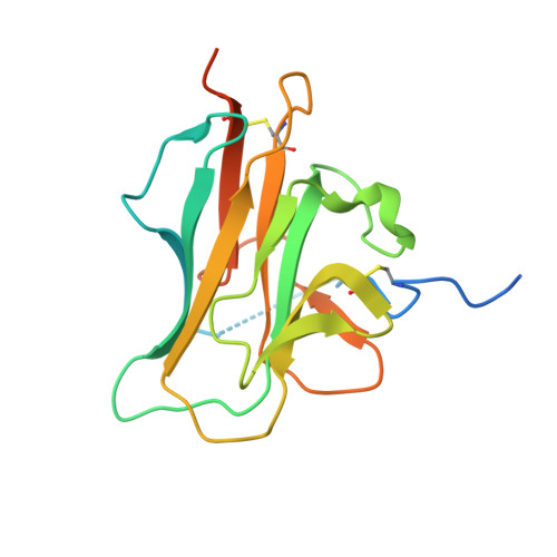
The crystal structure of the heparin-binding reelin-N domain of f-spondin.

Tan, K.Duquette, M.Liu, J.H.Lawler, J.Wang, J.H.

(2008) J Mol Biol 381: 1213-1223

  • DOI: https://doi.org/10.1016/j.jmb.2008.06.045
  • Primary Citation of Related Structures:  
    3COO

  • PubMed Abstract: 

    The extracellular matrix protein F-spondin mediates axon guidance during neuronal development. Its N-terminal domain, termed the reelin-N domain, is conserved in F-spondins, reelins, and other extracellular matrix proteins. In this study, a recombinant human reelin-N domain has been expressed, purified, and shown to bind heparin. The crystal structure of the reelin-N domain resolved to 2.0 A reveals a variant immunoglobulin-like fold and potential heparin-binding sites. Substantial conformational variations even in secondary structure are observed between the two chemically identical reelin-N domains in one crystallographic asymmetric unit. The variations may result from extensive, highly specific interactions across the interface of the two reelin-N domains. The calculated values of buried surface area and the interface's shape complementarity are consistent with the formation of a weak dimer. The homophilic asymmetric dimer can potentially offer advantages in binding to ligands such as glycosaminoglycans, which may, in turn, bridge the two reelin-N domains and stabilize the dimer.


  • Organizational Affiliation

    Department of Medical Oncology, Dana-Farber Cancer Institute, Boston, MA 02115, USA. ktan@anl.gov


Macromolecules
Find similar proteins by:  (by identity cutoff)  |  3D Structure
Entity ID: 1
MoleculeChains Sequence LengthOrganismDetailsImage
Spondin-1
A, B
180Homo sapiensMutation(s): 0 
Gene Names: Homo sapiens
UniProt & NIH Common Fund Data Resources
Find proteins for Q9HCB6 (Homo sapiens)
Explore Q9HCB6 
Go to UniProtKB:  Q9HCB6
PHAROS:  Q9HCB6
GTEx:  ENSG00000262655 
Entity Groups  
Sequence Clusters30% Identity50% Identity70% Identity90% Identity95% Identity100% Identity
UniProt GroupQ9HCB6
Sequence Annotations
Expand
  • Reference Sequence
Experimental Data & Validation

Experimental Data

  • Method: X-RAY DIFFRACTION
  • Resolution: 2.00 Å
  • R-Value Free: 0.241 
  • R-Value Work: 0.206 
  • R-Value Observed: 0.208 
  • Space Group: P 43 21 2
Unit Cell:
Length ( Å )Angle ( ˚ )
a = 58.605α = 90
b = 58.605β = 90
c = 219.542γ = 90
Software Package:
Software NamePurpose
REFMACrefinement
SBC-Collectdata collection
HKL-2000data reduction
HKL-2000data scaling
SHELXDphasing
MLPHAREphasing
DMphasing
RESOLVEphasing

Structure Validation

View Full Validation Report


Entry History 

Deposition Data

Revision History  (Full details and data files)

  • Version 1.0: 2008-08-26
    Type: Initial release
  • Version 1.1: 2011-07-13
    Changes: Advisory, Version format compliance
  • Version 1.2: 2017-10-25
    Changes: Refinement description