3COH

Crystal structure of Aurora-A in complex with a pentacyclic inhibitor


Experimental Data Snapshot

  • Method: X-RAY DIFFRACTION
  • Resolution: 2.70 Å
  • R-Value Free: 0.280 
  • R-Value Work: 0.246 
  • R-Value Observed: 0.248 

wwPDB Validation   3D Report Full Report


Ligand Structure Quality Assessment 


This is version 1.3 of the entry. See complete history


Literature

A pentacyclic aurora kinase inhibitor (AKI-001) with high in vivo potency and oral bioavailability.

Rawson, T.E.Ruth, M.Blackwood, E.Burdick, D.Corson, L.Dotson, J.Drummond, J.Fields, C.Georges, G.J.Goller, B.Halladay, J.Hunsaker, T.Kleinheinz, T.Krell, H.W.Li, J.Liang, J.Limberg, A.McNutt, A.Moffat, J.Phillips, G.Ran, Y.Safina, B.Ultsch, M.Walker, L.Wiesmann, C.Zhang, B.Zhou, A.Zhu, B.Y.Ruger, P.Cochran, A.G.

(2008) J Med Chem 51: 4465-4475

  • DOI: https://doi.org/10.1021/jm800052b
  • Primary Citation of Related Structures:  
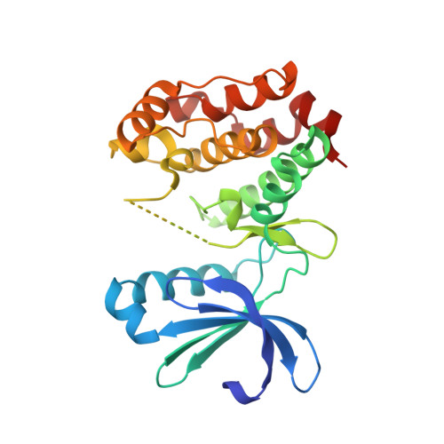
    3COH

  • PubMed Abstract: 

    Aurora kinase inhibitors have attracted a great deal of interest as a new class of antimitotic agents. We report a novel class of Aurora inhibitors based on a pentacyclic scaffold. A prototype pentacyclic inhibitor 32 (AKI-001) derived from two early lead structures improves upon the best properties of each parent and compares favorably to a previously reported Aurora inhibitor, 39 (VX-680). The inhibitor exhibits low nanomolar potency against both Aurora A and Aurora B enzymes, excellent cellular potency (IC50 < 100 nM), and good oral bioavailability. Phenotypic cellular assays show that both Aurora A and Aurora B are inhibited at inhibitor concentrations sufficient to block proliferation. Importantly, the cellular activity translates to potent inhibition of tumor growth in vivo. An oral dose of 5 mg/kg QD is well tolerated and results in near stasis (92% TGI) in an HCT116 mouse xenograft model.


  • Organizational Affiliation

    Departments of Small Molecule Drug DiscoVery, Cell Cycle and Global Regulators, Translational Oncology, and Protein Engineering, Genentech, Inc, 1 DNA Way, South San Francisco, California 94080, USA.


Macromolecules
Find similar proteins by:  (by identity cutoff)  |  3D Structure
Entity ID: 1
MoleculeChains Sequence LengthOrganismDetailsImage
Serine/threonine-protein kinase 6
A, B
268Homo sapiensMutation(s): 7 
Gene Names: AURKAAIKARK1AURABTAKSTK15STK6
EC: 2.7.11.1
UniProt & NIH Common Fund Data Resources
Find proteins for O14965 (Homo sapiens)
Explore O14965 
Go to UniProtKB:  O14965
PHAROS:  O14965
GTEx:  ENSG00000087586 
Entity Groups  
Sequence Clusters30% Identity50% Identity70% Identity90% Identity95% Identity100% Identity
UniProt GroupO14965
Sequence Annotations
Expand
  • Reference Sequence
Small Molecules
Ligands 1 Unique
IDChains Name / Formula / InChI Key2D Diagram3D Interactions
83H
Query on 83H

Download Ideal Coordinates CCD File 
C [auth A],
D [auth B]
8-ethyl-3,10,10-trimethyl-4,5,6,8,10,12-hexahydropyrazolo[4',3':6,7]cyclohepta[1,2-b]pyrrolo[2,3-f]indol-9(1H)-one
C21 H24 N4 O
AOMMPEGZDRAGRC-UHFFFAOYSA-N
Binding Affinity Annotations 
IDSourceBinding Affinity
83H PDBBind:  3COH IC50: 4 (nM) from 1 assay(s)
Experimental Data & Validation

Experimental Data

  • Method: X-RAY DIFFRACTION
  • Resolution: 2.70 Å
  • R-Value Free: 0.280 
  • R-Value Work: 0.246 
  • R-Value Observed: 0.248 
  • Space Group: P 42 21 2
Unit Cell:
Length ( Å )Angle ( ˚ )
a = 99.542α = 90
b = 99.542β = 90
c = 138.845γ = 90
Software Package:
Software NamePurpose
REFMACrefinement
Blu-Icedata collection
HKL-2000data reduction
SCALEPACKdata scaling
AMoREphasing

Structure Validation

View Full Validation Report


Ligand Structure Quality Assessment 


Entry History 

Deposition Data

Revision History  (Full details and data files)

  • Version 1.0: 2009-02-17
    Type: Initial release
  • Version 1.1: 2011-07-13
    Changes: Advisory, Refinement description, Version format compliance
  • Version 1.2: 2021-10-20
    Changes: Database references, Derived calculations
  • Version 1.3: 2023-08-30
    Changes: Data collection, Refinement description