3CDZ

Crystal structure of human factor VIII


Experimental Data Snapshot

  • Method: X-RAY DIFFRACTION
  • Resolution: 3.98 Å
  • R-Value Free: 0.327 
  • R-Value Work: 0.256 
  • R-Value Observed: 0.259 

wwPDB Validation   3D Report Full Report


Ligand Structure Quality Assessment 


This is version 2.1 of the entry. See complete history


Literature

Crystal structure of human factor VIII: implications for the formation of the factor IXa-factor VIIIa complex.

Ngo, J.C.Huang, M.Roth, D.A.Furie, B.C.Furie, B.

(2008) Structure 16: 597-606

  • DOI: https://doi.org/10.1016/j.str.2008.03.001
  • Primary Citation of Related Structures:  
    3CDZ

  • PubMed Abstract: 

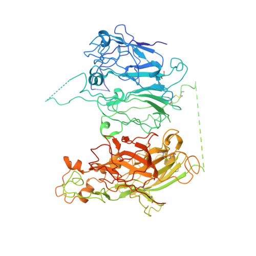
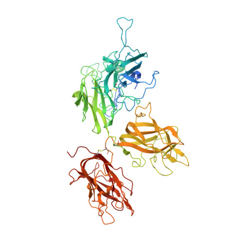
    Factor VIII is a procofactor that plays a critical role in blood coagulation, and is missing or defective in hemophilia A. We determined the X-ray crystal structure of B domain-deleted human factor VIII. This protein is composed of five globular domains and contains one Ca(2+) and two Cu(2+) ions. The three homologous A domains form a triangular heterotrimer where the A1 and A3 domains serve as the base and interact with the C2 and C1 domains, respectively. The structurally homologous C1 and C2 domains reveal membrane binding features. Based on biochemical studies, a model of the factor IXa-factor VIIIa complex was constructed by in silico docking. Factor IXa wraps across the side of factor VIII, and an extended interface spans the factor VIII heavy and light chains. This model provides insight into the activation of factor VIII and the interaction of factor VIIIa with factor IXa on the membrane surface.


  • Organizational Affiliation

    Marine Biological Laboratory, Woods Hole, MA 02543, USA.


Macromolecules
Find similar proteins by:  (by identity cutoff)  |  3D Structure
Entity ID: 1
MoleculeChains Sequence LengthOrganismDetailsImage
Coagulation factor VIII heavy chain754Homo sapiensMutation(s): 0 
Gene Names: F8F8C
UniProt & NIH Common Fund Data Resources
Find proteins for P00451 (Homo sapiens)
Explore P00451 
Go to UniProtKB:  P00451
PHAROS:  P00451
GTEx:  ENSG00000185010 
Entity Groups  
Sequence Clusters30% Identity50% Identity70% Identity90% Identity95% Identity100% Identity
UniProt GroupP00451
Sequence Annotations
Expand
  • Reference Sequence
Find similar proteins by:  (by identity cutoff)  |  3D Structure
Entity ID: 2
MoleculeChains Sequence LengthOrganismDetailsImage
Coagulation factor VIII light chain684Homo sapiensMutation(s): 1 
Gene Names: F8F8C
UniProt & NIH Common Fund Data Resources
Find proteins for P00451 (Homo sapiens)
Explore P00451 
Go to UniProtKB:  P00451
PHAROS:  P00451
GTEx:  ENSG00000185010 
Entity Groups  
Sequence Clusters30% Identity50% Identity70% Identity90% Identity95% Identity100% Identity
UniProt GroupP00451
Sequence Annotations
Expand
  • Reference Sequence
Oligosaccharides

Help

Entity ID: 3
MoleculeChains Length2D Diagram Glycosylation3D Interactions
2-acetamido-2-deoxy-beta-D-glucopyranose-(1-4)-2-acetamido-2-deoxy-beta-D-glucopyranose
C
2N-Glycosylation
Glycosylation Resources
GlyTouCan:  G42666HT
GlyCosmos:  G42666HT
GlyGen:  G42666HT
Entity ID: 4
MoleculeChains Length2D Diagram Glycosylation3D Interactions
alpha-D-mannopyranose-(1-3)-[alpha-D-mannopyranose-(1-6)]alpha-D-mannopyranose-(1-4)-2-acetamido-2-deoxy-beta-D-glucopyranose
D
4N/A
Glycosylation Resources
GlyTouCan:  G95760DN
GlyCosmos:  G95760DN
GlyGen:  G95760DN
Experimental Data & Validation

Experimental Data

  • Method: X-RAY DIFFRACTION
  • Resolution: 3.98 Å
  • R-Value Free: 0.327 
  • R-Value Work: 0.256 
  • R-Value Observed: 0.259 
  • Space Group: P 41 21 2
Unit Cell:
Length ( Å )Angle ( ˚ )
a = 134.113α = 90
b = 134.113β = 90
c = 349.76γ = 90
Software Package:
Software NamePurpose
DENZOdata reduction
SCALEPACKdata scaling
AMoREphasing
REFMACrefinement
PDB_EXTRACTdata extraction
HKL-2000data collection
HKL-2000data reduction
HKL-2000data scaling

Structure Validation

View Full Validation Report


Ligand Structure Quality Assessment 


Entry History 

Deposition Data

Revision History  (Full details and data files)

  • Version 1.0: 2008-04-01
    Type: Initial release
  • Version 1.1: 2011-07-13
    Changes: Advisory, Version format compliance
  • Version 1.2: 2017-10-25
    Changes: Advisory, Refinement description
  • Version 2.0: 2020-07-29
    Type: Remediation
    Reason: Carbohydrate remediation
    Changes: Advisory, Atomic model, Data collection, Database references, Derived calculations, Structure summary
  • Version 2.1: 2023-08-30
    Changes: Data collection, Database references, Refinement description, Structure summary