The 1.25 A resolution structure of phosphoribosyl-ATP pyrophosphohydrolase from Mycobacterium tuberculosis.
Javid-Majd, F., Yang, D., Ioerger, T.R., Sacchettini, J.C.(2008) Acta Crystallogr D Biol Crystallogr 64: 627-635
- PubMed: 18560150
- DOI: https://doi.org/10.1107/S0907444908007105
- Primary Citation of Related Structures:
1Y6X, 3C90 - PubMed Abstract:
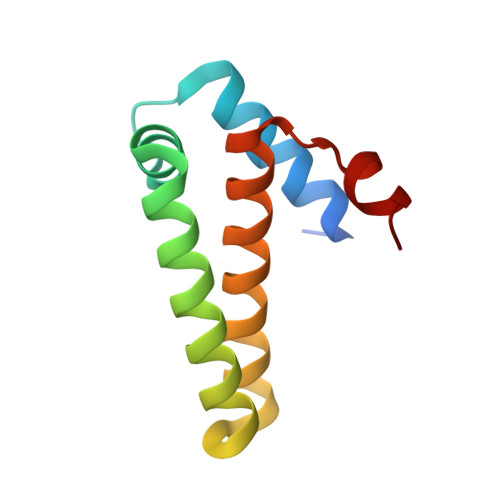
Phosphoribosyl-ATP pyrophosphohydrolase is the second enzyme in the histidine-biosynthetic pathway, irreversibly hydrolyzing phosphoribosyl-ATP to phosphoribosyl-AMP and pyrophosphate. It is encoded by the hisE gene, which is present as a separate gene in many bacteria and archaea but is fused to hisI in other bacteria, fungi and plants. Because of its essentiality for growth in vitro, HisE is a potential drug target for tuberculosis. The crystal structures of two native (uncomplexed) forms of HisE from Mycobacterium tuberculosis have been determined to resolutions of 1.25 and 1.79 A. The structure of the apoenzyme reveals that the protein is composed of five alpha-helices with connecting loops and is a member of the alpha-helical nucleoside-triphosphate pyrophosphatase superfamily. The biological unit of the protein is a homodimer, with an active site on each subunit composed of residues exclusively from that subunit. A comparison with the Campylobacter jejuni dUTPase active site allowed the identification of putative metal- and substrate-binding sites in HisE, including four conserved glutamate and glutamine residues in the sequence that are consistent with a motif for pyrophosphohydrolase activity. However, significant differences between family members are observed in the loop region between alpha-helices H1 and H3. The crystal structure of M. tuberculosis HisE provides insights into possible mechanisms of substrate binding and the diversity of the nucleoside-triphosphate pyrophosphatase superfamily.
Organizational Affiliation:
Department of Biochemistry and Biophysics, Texas A&M University, College Station, Texas 77843-2128, USA.