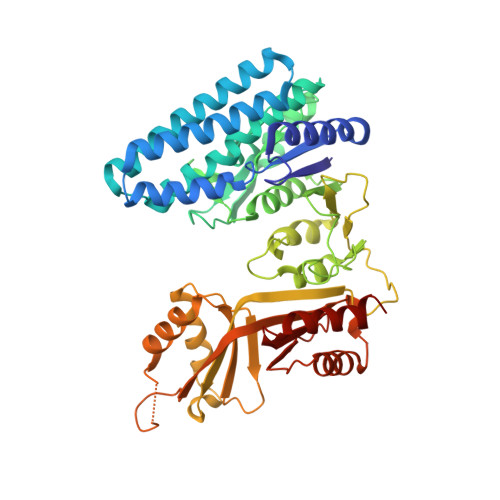
The Structural Basis for Allosteric Inhibition of a Threonine-sensitive Aspartokinase.
Liu, X., Pavlovsky, A.G., Viola, R.E.(2008) J Biol Chem 283: 16216-16225
- PubMed: 18334478
- DOI: https://doi.org/10.1074/jbc.M800760200
- Primary Citation of Related Structures:
3C1M, 3C1N, 3C20 - PubMed Abstract:
The commitment step to the aspartate pathway of amino acid biosynthesis is the phosphorylation of aspartic acid catalyzed by aspartokinase (AK). Most microorganisms and plants have multiple forms of this enzyme, and many of these isofunctional enzymes are subject to feedback regulation by the end products of the pathway. However, the archeal species Methanococcus jannaschii has only a single, monofunctional form of AK. The substrate l-aspartate binds to this recombinant enzyme in two different orientations, providing the first structural evidence supporting the relaxed regiospecificity previously observed with several alternative substrates of Escherichia coli AK ( Angeles, T. S., Hunsley, J. R., and Viola, R. E. (1992) Biochemistry 31, 799-805 ). Binding of the nucleotide substrate triggers significant domain movements that result in a more compact quaternary structure. In contrast, the highly cooperative binding of the allosteric regulator l-threonine to multiple sites on this dimer of dimers leads to an open enzyme structure. A comparison of these structures supports a mechanism for allosteric regulation in which the domain movements induced by threonine binding causes displacement of the substrates from the enzyme, resulting in a relaxed, inactive conformation.
Organizational Affiliation:
Department of Chemistry, University of Toledo, Toledo, Ohio 43606.