3BT2

Structure of urokinase receptor, urokinase and vitronectin complex


Experimental Data Snapshot

  • Method: X-RAY DIFFRACTION
  • Resolution: 2.50 Å
  • R-Value Free: 0.272 
  • R-Value Work: 0.229 
  • R-Value Observed: 0.230 

wwPDB Validation   3D Report Full Report


Ligand Structure Quality Assessment 


This is version 2.1 of the entry. See complete history


Literature

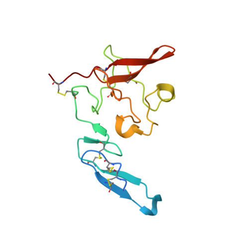
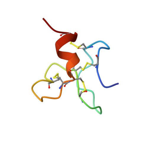
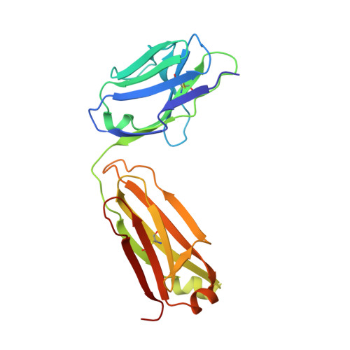
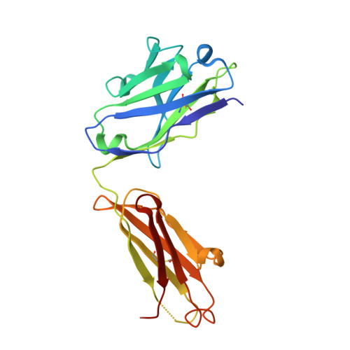
Crystal structures of two human vitronectin, urokinase and urokinase receptor complexes

Huai, Q.Zhou, A.Lin, L.Mazar, A.P.Parry, G.C.Callahan, J.Shaw, D.E.Furie, B.Furie, B.C.Huang, M.

(2008) Nat Struct Mol Biol 15: 422-423

  • DOI: https://doi.org/10.1038/nsmb.1404
  • Primary Citation of Related Structures:  
    3BT1, 3BT2

  • PubMed Abstract: 

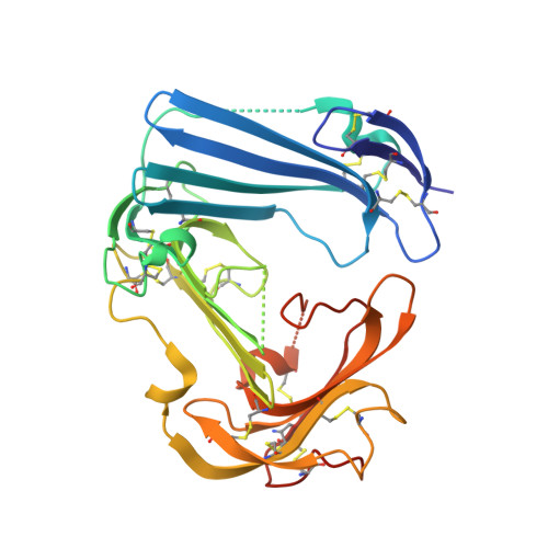
    The urokinase receptor (uPAR) can recognize several ligands. The structural basis for this multiple ligand recognition by uPAR is unknown. This study reports the crystal structures of uPAR in complex with both urokinase (uPA) and vitronectin and reveal that uPA occupies the central cavity of the receptor, whereas vitronectin binds at the outer side of the receptor. These results provide a structural understanding of one receptor binding to two ligands.


  • Organizational Affiliation

    Division of Hemostasis and Thrombosis, Beth Israel Deaconess Medical Center and Harvard Medical School, Boston, Massachusetts 02215, USA.


Macromolecules
Find similar proteins by:  (by identity cutoff)  |  3D Structure
Entity ID: 1
MoleculeChains Sequence LengthOrganismDetailsImage
Urokinase-type plasminogen activator135Homo sapiensMutation(s): 0 
Gene Names: PLAU
UniProt & NIH Common Fund Data Resources
Find proteins for P00749 (Homo sapiens)
Explore P00749 
Go to UniProtKB:  P00749
PHAROS:  P00749
GTEx:  ENSG00000122861 
Entity Groups  
Sequence Clusters30% Identity50% Identity70% Identity90% Identity95% Identity100% Identity
UniProt GroupP00749
Sequence Annotations
Expand
  • Reference Sequence
Find similar proteins by:  (by identity cutoff)  |  3D Structure
Entity ID: 2
MoleculeChains Sequence LengthOrganismDetailsImage
Vitronectin40Homo sapiensMutation(s): 0 
Gene Names: VTN
UniProt & NIH Common Fund Data Resources
Find proteins for P04004 (Homo sapiens)
Explore P04004 
Go to UniProtKB:  P04004
PHAROS:  P04004
GTEx:  ENSG00000109072 
Entity Groups  
Sequence Clusters30% Identity50% Identity70% Identity90% Identity95% Identity100% Identity
UniProt GroupP04004
Sequence Annotations
Expand
  • Reference Sequence
Find similar proteins by:  (by identity cutoff)  |  3D Structure
Entity ID: 3
MoleculeChains Sequence LengthOrganismDetailsImage
anti-uPAR antibody, light chainC [auth L]212Mus musculusMutation(s): 0 
Entity Groups  
Sequence Clusters30% Identity50% Identity70% Identity90% Identity95% Identity100% Identity
Sequence Annotations
Expand
  • Reference Sequence
Find similar proteins by:  (by identity cutoff)  |  3D Structure
Entity ID: 4
MoleculeChains Sequence LengthOrganismDetailsImage
anti-uPAR antibody, heavy chainD [auth H]214Mus musculusMutation(s): 0 
Entity Groups  
Sequence Clusters30% Identity50% Identity70% Identity90% Identity95% Identity100% Identity
Sequence Annotations
Expand
  • Reference Sequence
Find similar proteins by:  (by identity cutoff)  |  3D Structure
Entity ID: 5
MoleculeChains Sequence LengthOrganismDetailsImage
Urokinase plasminogen activator surface receptorE [auth U]283Homo sapiensMutation(s): 0 
Gene Names: PLAURMO3UPAR
UniProt & NIH Common Fund Data Resources
Find proteins for Q03405 (Homo sapiens)
Explore Q03405 
Go to UniProtKB:  Q03405
PHAROS:  Q03405
GTEx:  ENSG00000011422 
Entity Groups  
Sequence Clusters30% Identity50% Identity70% Identity90% Identity95% Identity100% Identity
UniProt GroupQ03405
Sequence Annotations
Expand
  • Reference Sequence
Oligosaccharides

Help

Entity ID: 6
MoleculeChains Length2D Diagram Glycosylation3D Interactions
2-acetamido-2-deoxy-beta-D-glucopyranose-(1-4)-2-acetamido-2-deoxy-beta-D-glucopyranoseF [auth C],
G [auth D]
2N-Glycosylation
Glycosylation Resources
GlyTouCan:  G42666HT
GlyCosmos:  G42666HT
GlyGen:  G42666HT
Small Molecules
Ligands 1 Unique
IDChains Name / Formula / InChI Key2D Diagram3D Interactions
NAG
Query on NAG

Download Ideal Coordinates CCD File 
H [auth U]2-acetamido-2-deoxy-beta-D-glucopyranose
C8 H15 N O6
OVRNDRQMDRJTHS-FMDGEEDCSA-N
Experimental Data & Validation

Experimental Data

  • Method: X-RAY DIFFRACTION
  • Resolution: 2.50 Å
  • R-Value Free: 0.272 
  • R-Value Work: 0.229 
  • R-Value Observed: 0.230 
  • Space Group: P 1 21 1
Unit Cell:
Length ( Å )Angle ( ˚ )
a = 52.096α = 90
b = 87.205β = 94.31
c = 124.274γ = 90
Software Package:
Software NamePurpose
REFMACrefinement
HKL-2000data collection
HKL-2000data reduction
HKL-2000data scaling
AMoREphasing

Structure Validation

View Full Validation Report


Ligand Structure Quality Assessment 


Entry History 

Deposition Data

  • Released Date: 2008-03-25 
  • Deposition Author(s): Huang, M.

Revision History  (Full details and data files)

  • Version 1.0: 2008-03-25
    Type: Initial release
  • Version 1.1: 2011-07-13
    Changes: Advisory, Version format compliance
  • Version 2.0: 2020-07-29
    Type: Remediation
    Reason: Carbohydrate remediation
    Changes: Advisory, Atomic model, Data collection, Database references, Derived calculations, Structure summary
  • Version 2.1: 2023-11-01
    Changes: Data collection, Database references, Refinement description, Structure summary