3BP8

Crystal structure of Mlc/EIIB complex


Experimental Data Snapshot

  • Method: X-RAY DIFFRACTION
  • Resolution: 2.85 Å
  • R-Value Free: 0.301 
  • R-Value Work: 0.228 
  • R-Value Observed: 0.228 

wwPDB Validation   3D Report Full Report


This is version 1.2 of the entry. See complete history


Literature

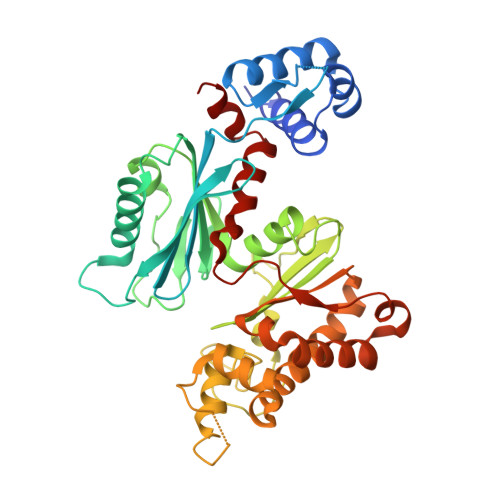
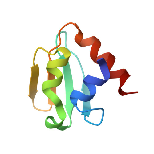
Analyses of Mlc-IIBGlc interaction and a plausible molecular mechanism of Mlc inactivation by membrane sequestration

Nam, T.W.Jung, H.I.An, Y.J.Park, Y.H.Lee, S.H.Seok, Y.J.Cha, S.S.

(2008) Proc Natl Acad Sci U S A 105: 3751-3756

  • DOI: https://doi.org/10.1073/pnas.0709295105
  • Primary Citation of Related Structures:  
    3BP3, 3BP8

  • PubMed Abstract: 

    In Escherichia coli, glucose-dependent transcriptional induction of genes encoding a variety of sugar-metabolizing enzymes and transport systems is mediated by the phosphorylation state-dependent interaction of membrane-bound enzyme IICB(Glc) (EIICB(Glc)) with the global repressor Mlc. Here we report the crystal structure of a tetrameric Mlc in a complex with four molecules of enzyme IIB(Glc) (EIIB), the cytoplasmic domain of EIICB(Glc). Each monomer of Mlc has one bound EIIB molecule, indicating the 1:1 stoichiometry. The detailed view of the interface, along with the high-resolution structure of EIIB containing a sulfate ion at the phosphorylation site, suggests that the phosphorylation-induced steric hindrance and disturbance of polar intermolecular interactions impede complex formation. Furthermore, we reveal that Mlc possesses a built-in flexibility for the structural adaptation to its target DNA and that interaction of Mlc with EIIB fused only to dimeric proteins resulted in the loss of its DNA binding ability, suggesting that flexibility of the Mlc structure is indispensable for its DNA binding.


  • Organizational Affiliation

    Department of Biological Sciences and Institute of Microbiology, Seoul National University, Seoul 151-742, Korea.


Macromolecules
Find similar proteins by:  (by identity cutoff)  |  3D Structure
Entity ID: 1
MoleculeChains Sequence LengthOrganismDetailsImage
Putative NAGC-like transcriptional regulator
A, B
406Escherichia coliMutation(s): 0 
Gene Names: mlc
UniProt
Find proteins for P50456 (Escherichia coli (strain K12))
Explore P50456 
Go to UniProtKB:  P50456
Entity Groups  
Sequence Clusters30% Identity50% Identity70% Identity90% Identity95% Identity100% Identity
UniProt GroupP50456
Sequence Annotations
Expand
  • Reference Sequence
Find similar proteins by:  (by identity cutoff)  |  3D Structure
Entity ID: 2
MoleculeChains Sequence LengthOrganismDetailsImage
PTS system glucose-specific EIICB component
C, D
75Escherichia coliMutation(s): 0 
Gene Names: ptsGglcAumg
UniProt
Find proteins for P69786 (Escherichia coli (strain K12))
Explore P69786 
Go to UniProtKB:  P69786
Entity Groups  
Sequence Clusters30% Identity50% Identity70% Identity90% Identity95% Identity100% Identity
UniProt GroupP69786
Sequence Annotations
Expand
  • Reference Sequence
Experimental Data & Validation

Experimental Data

  • Method: X-RAY DIFFRACTION
  • Resolution: 2.85 Å
  • R-Value Free: 0.301 
  • R-Value Work: 0.228 
  • R-Value Observed: 0.228 
  • Space Group: C 1 2 1
Unit Cell:
Length ( Å )Angle ( ˚ )
a = 201.427α = 90
b = 55.424β = 95.29
c = 82.46γ = 90
Software Package:
Software NamePurpose
PROTEUM PLUSdata collection
MOLREPphasing
CNSrefinement
HKL-2000data reduction
HKL-2000data scaling

Structure Validation

View Full Validation Report


Entry History 

Deposition Data

Revision History  (Full details and data files)

  • Version 1.0: 2008-05-27
    Type: Initial release
  • Version 1.1: 2011-07-13
    Changes: Version format compliance
  • Version 1.2: 2023-11-01
    Changes: Data collection, Database references, Derived calculations, Refinement description