3BNG

W. succinogenes NrfA Y218F


Experimental Data Snapshot

  • Method: X-RAY DIFFRACTION
  • Resolution: 1.50 Å
  • R-Value Free: 0.215 
  • R-Value Work: 0.181 
  • R-Value Observed: 0.183 

wwPDB Validation   3D Report Full Report


Ligand Structure Quality Assessment 


This is version 1.3 of the entry. See complete history


Literature

Binding and Reduction of Sulfite by Cytochrome c Nitrite Reductase

Lukat, P.Rudolf, M.Stach, P.Messerschmidt, A.Kroneck, P.M.H.Simon, J.Einsle, O.

(2008) Biochemistry 47: 2080-2086

  • DOI: https://doi.org/10.1021/bi7021415
  • Primary Citation of Related Structures:  
    3BNF, 3BNG, 3BNH, 3BNJ

  • PubMed Abstract: 

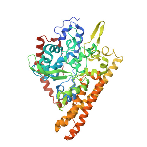
    Pentaheme cytochrome c nitrite reductase (ccNiR) catalyzes the six-electron reduction of nitrite to ammonia as the final step in the dissimilatory pathway of nitrate ammonification. It has also been shown to reduce sulfite to sulfide, thus forming the only known link between the biogeochemical cycles of nitrogen and of sulfur. We have found the sulfite reductase activity of ccNiR from Wolinella succinogenes to be significantly smaller than its nitrite reductase activity but still several times higher than the one described for dissimilatory, siroheme-containing sulfite reductases. To compare the sulfite reductase activity of ccNiR with our previous data on nitrite reduction, we determined the binding mode of sulfite to the catalytic heme center of ccNiR from W. succinogenes at a resolution of 1.7 A. Sulfite and nitrite both provide a pair of electrons to form the coordinative bond to the Fe(III) active site of the enzyme, and the oxygen atoms of sulfite are found to interact with the three active site protein residues conserved within the enzyme family. Furthermore, we have characterized the active site variant Y218F of ccNiR that exhibited an almost complete loss of nitrite reductase activity, while sulfite reduction remained unaffected. These data provide a first direct insight into the role of the first sphere of protein ligands at the active site in ccNiR catalysis.


  • Organizational Affiliation

    Institut für Mikrobiologie und Genetik, Abteilung Molekulare Strukturbiologie, Georg-August-Universität Göttingen, 37077 Göttingen, Germany.


Macromolecules
Find similar proteins by:  (by identity cutoff)  |  3D Structure
Entity ID: 1
MoleculeChains Sequence LengthOrganismDetailsImage
Cytochrome c-552485Wolinella succinogenesMutation(s): 1 
Gene Names: nrfA Y218F
EC: 1.7.2.2
UniProt
Find proteins for Q9S1E5 (Wolinella succinogenes (strain ATCC 29543 / DSM 1740 / LMG 7466 / NCTC 11488 / FDC 602W))
Explore Q9S1E5 
Go to UniProtKB:  Q9S1E5
Entity Groups  
Sequence Clusters30% Identity50% Identity70% Identity90% Identity95% Identity100% Identity
UniProt GroupQ9S1E5
Sequence Annotations
Expand
  • Reference Sequence
Small Molecules
Ligands 5 Unique
IDChains Name / Formula / InChI Key2D Diagram3D Interactions
HEM
Query on HEM

Download Ideal Coordinates CCD File 
L [auth A],
M [auth A],
N [auth A],
O [auth A],
P [auth A]
PROTOPORPHYRIN IX CONTAINING FE
C34 H32 Fe N4 O4
KABFMIBPWCXCRK-RGGAHWMASA-L
SO4
Query on SO4

Download Ideal Coordinates CCD File 
E [auth A],
F [auth A]
SULFATE ION
O4 S
QAOWNCQODCNURD-UHFFFAOYSA-L
Y1
Query on Y1

Download Ideal Coordinates CCD File 
H [auth A],
I [auth A],
J [auth A],
K [auth A]
YTTRIUM ION
Y
KAJPZYFHSCFBCI-UHFFFAOYSA-N
ACT
Query on ACT

Download Ideal Coordinates CCD File 
B [auth A],
C [auth A],
D [auth A]
ACETATE ION
C2 H3 O2
QTBSBXVTEAMEQO-UHFFFAOYSA-M
CA
Query on CA

Download Ideal Coordinates CCD File 
G [auth A]CALCIUM ION
Ca
BHPQYMZQTOCNFJ-UHFFFAOYSA-N
Experimental Data & Validation

Experimental Data

  • Method: X-RAY DIFFRACTION
  • Resolution: 1.50 Å
  • R-Value Free: 0.215 
  • R-Value Work: 0.181 
  • R-Value Observed: 0.183 
  • Space Group: I 41 2 2
Unit Cell:
Length ( Å )Angle ( ˚ )
a = 120.03α = 90
b = 120.03β = 90
c = 186.276γ = 90
Software Package:
Software NamePurpose
REFMACrefinement
MAR345dtbdata collection
DENZOdata reduction
SCALEPACKdata scaling
MOLREPphasing

Structure Validation

View Full Validation Report


Ligand Structure Quality Assessment 


Entry History 

Deposition Data

Revision History  (Full details and data files)

  • Version 1.0: 2008-02-26
    Type: Initial release
  • Version 1.1: 2011-07-13
    Changes: Version format compliance
  • Version 1.2: 2021-11-10
    Changes: Data collection, Database references, Derived calculations
  • Version 1.3: 2023-11-01
    Changes: Data collection, Refinement description