3BL8

Crystal structure of the extracellular domain of neuroligin 2A from mouse


Experimental Data Snapshot

  • Method: X-RAY DIFFRACTION
  • Resolution: 3.30 Å
  • R-Value Free: 0.262 
  • R-Value Work: 0.219 
  • R-Value Observed: 0.221 

wwPDB Validation   3D Report Full Report


Ligand Structure Quality Assessment 


This is version 2.1 of the entry. See complete history


Literature

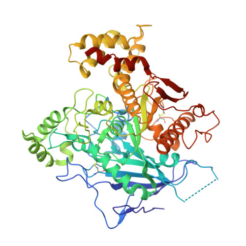
Crystal structure of the extracellular cholinesterase-like domain from neuroligin-2.

Koehnke, J.Jin, X.Budreck, E.C.Posy, S.Scheiffele, P.Honig, B.Shapiro, L.

(2008) Proc Natl Acad Sci U S A 105: 1873-1878

  • DOI: https://doi.org/10.1073/pnas.0711701105
  • Primary Citation of Related Structures:  
    3BL8

  • PubMed Abstract: 

    Neuroligins (NLs) are catalytically inactive members of a family of cholinesterase-like transmembrane proteins that mediate cell adhesion at neuronal synapses. Postsynaptic neuroligins engage in Ca2+-dependent transsynaptic interactions via their extracellular cholinesterase domain with presynaptic neurexins (NRXs). These interactions may be regulated by two short splice insertions (termed A and B) in the NL cholinesterase domain. Here, we present the 3.3-A crystal structure of the ectodomain from NL2 containing splice insertion A (NL2A). The overall structure of NL2A resembles that of cholinesterases, but several structural features are unique to the NL proteins. First, structural elements surrounding the esterase active-site region differ significantly between active esterases and NL2A. On the opposite surface of the NL2A molecule, the positions of the A and B splice insertions identify a candidate NRX interaction site of the NL protein. Finally, sequence comparisons of NL isoforms allow for mapping the location of residues of previously identified mutations in NL3 and NL4 found in patients with autism spectrum disorders. Overall, the NL2 structure promises to provide a valuable model for dissecting NL isoform- and synapse-specific functions.


  • Organizational Affiliation

    Department of Biochemistry and Molecular Biophysics, Columbia University, New York, NY 10032, USA.


Macromolecules
Find similar proteins by:  (by identity cutoff)  |  3D Structure
Entity ID: 1
MoleculeChains Sequence LengthOrganismDetailsImage
Neuroligin-2
A, B, C, D
580Mus musculusMutation(s): 0 
Gene Names: Nlgn2Kiaa1366
Membrane Entity: Yes 
UniProt
Find proteins for Q69ZK9 (Mus musculus)
Explore Q69ZK9 
Go to UniProtKB:  Q69ZK9
Entity Groups  
Sequence Clusters30% Identity50% Identity70% Identity90% Identity95% Identity100% Identity
UniProt GroupQ69ZK9
Sequence Annotations
Expand
  • Reference Sequence
Oligosaccharides

Help

Entity ID: 2
MoleculeChains Length2D Diagram Glycosylation3D Interactions
alpha-D-mannopyranose-(1-4)-2-acetamido-2-deoxy-beta-D-glucopyranose-(1-4)-2-acetamido-2-deoxy-beta-D-glucopyranose
E
3N-Glycosylation
Glycosylation Resources
GlyTouCan:  G62182OO
GlyCosmos:  G62182OO
GlyGen:  G62182OO
Entity ID: 3
MoleculeChains Length2D Diagram Glycosylation3D Interactions
alpha-D-mannopyranose-(1-3)-[alpha-D-mannopyranose-(1-6)]alpha-D-mannopyranose-(1-3)-[alpha-D-mannopyranose-(1-6)]beta-D-mannopyranose-(1-4)-2-acetamido-2-deoxy-beta-D-glucopyranose-(1-4)-2-acetamido-2-deoxy-beta-D-glucopyranose
F
7N-Glycosylation
Glycosylation Resources
GlyTouCan:  G97423PY
GlyCosmos:  G97423PY
GlyGen:  G97423PY
Small Molecules
Ligands 1 Unique
IDChains Name / Formula / InChI Key2D Diagram3D Interactions
NAG
Query on NAG

Download Ideal Coordinates CCD File 
G [auth A],
H [auth B]
2-acetamido-2-deoxy-beta-D-glucopyranose
C8 H15 N O6
OVRNDRQMDRJTHS-FMDGEEDCSA-N
Experimental Data & Validation

Experimental Data

  • Method: X-RAY DIFFRACTION
  • Resolution: 3.30 Å
  • R-Value Free: 0.262 
  • R-Value Work: 0.219 
  • R-Value Observed: 0.221 
  • Space Group: C 1 2 1
Unit Cell:
Length ( Å )Angle ( ˚ )
a = 214.723α = 90
b = 92.567β = 98.36
c = 188.408γ = 90
Software Package:
Software NamePurpose
DENZOdata reduction
SCALEPACKdata scaling
PHASERphasing
DMphasing
REFMACrefinement
PDB_EXTRACTdata extraction
ADSCdata collection

Structure Validation

View Full Validation Report


Ligand Structure Quality Assessment 


Entry History 

Deposition Data

Revision History  (Full details and data files)

  • Version 1.0: 2008-02-19
    Type: Initial release
  • Version 1.1: 2011-07-13
    Changes: Advisory, Version format compliance
  • Version 1.2: 2017-10-25
    Changes: Advisory, Refinement description
  • Version 2.0: 2020-07-29
    Type: Remediation
    Reason: Carbohydrate remediation
    Changes: Advisory, Atomic model, Data collection, Database references, Derived calculations, Structure summary
  • Version 2.1: 2021-03-31
    Changes: Source and taxonomy, Structure summary