3BGF

X-ray crystal structure of the SARS coronavirus spike receptor binding domain in complex with F26G19 Fab


Experimental Data Snapshot

  • Method: X-RAY DIFFRACTION
  • Resolution: 3.00 Å
  • R-Value Free: 0.286 
  • R-Value Work: 0.234 
  • R-Value Observed: 0.237 

wwPDB Validation   3D Report Full Report


This is version 1.3 of the entry. See complete history


Literature

Structural insights into immune recognition of the severe acute respiratory syndrome coronavirus S protein receptor binding domain.

Pak, J.E.Sharon, C.Satkunarajah, M.Auperin, T.C.Cameron, C.M.Kelvin, D.J.Seetharaman, J.Cochrane, A.Plummer, F.A.Berry, J.D.Rini, J.M.

(2009) J Mol Biol 388: 815-823

  • DOI: https://doi.org/10.1016/j.jmb.2009.03.042
  • Primary Citation of Related Structures:  
    3BGF

  • PubMed Abstract: 

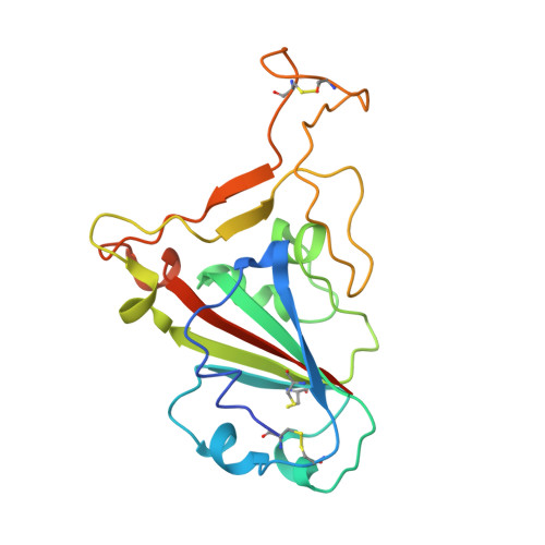
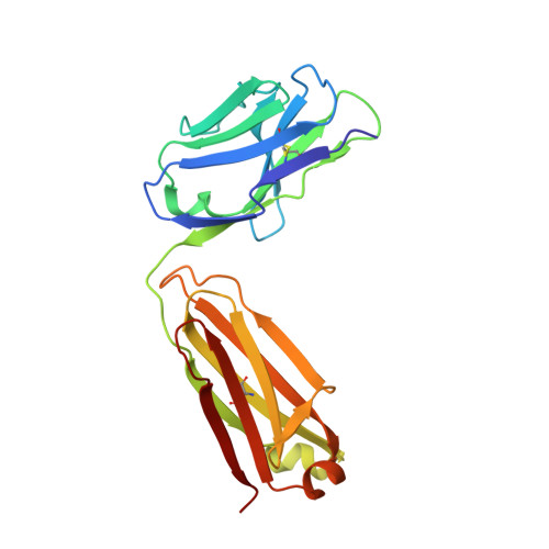
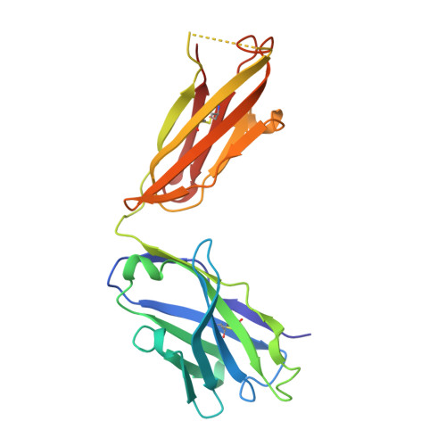
    The spike (S) protein of the severe acute respiratory syndrome coronavirus (SARS-CoV) is responsible for host cell attachment and fusion of the viral and host cell membranes. Within S the receptor binding domain (RBD) mediates the interaction with angiotensin-converting enzyme 2 (ACE2), the SARS-CoV host cell receptor. Both S and the RBD are highly immunogenic and both have been found to elicit neutralizing antibodies. Reported here is the X-ray crystal structure of the RBD in complex with the Fab of a neutralizing mouse monoclonal antibody, F26G19, elicited by immunization with chemically inactivated SARS-CoV. The RBD-F26G19 Fab complex represents the first example of the structural characterization of an antibody elicited by an immune response to SARS-CoV or any fragment of it. The structure reveals that the RBD surface recognized by F26G19 overlaps significantly with the surface recognized by ACE2 and, as such, suggests that F26G19 likely neutralizes SARS-CoV by blocking the virus-host cell interaction.


  • Organizational Affiliation

    Department of Molecular Genetics, University of Toronto, Toronto, Ontario, Canada.


Macromolecules
Find similar proteins by:  (by identity cutoff)  |  3D Structure
Entity ID: 1
MoleculeChains Sequence LengthOrganismDetailsImage
Spike protein S1A [auth S],
B [auth A]
193Severe acute respiratory syndrome-related coronavirusMutation(s): 0 
Gene Names: S
UniProt
Find proteins for P59594 (Severe acute respiratory syndrome coronavirus)
Explore P59594 
Go to UniProtKB:  P59594
Entity Groups  
Sequence Clusters30% Identity50% Identity70% Identity90% Identity95% Identity100% Identity
UniProt GroupP59594
Sequence Annotations
Expand
  • Reference Sequence
Find similar proteins by:  (by identity cutoff)  |  3D Structure
Entity ID: 2
MoleculeChains Sequence LengthOrganismDetailsImage
F26G19 FabC [auth L],
E [auth C]
212Mus musculusMutation(s): 0 
UniProt
Find proteins for Q7TS98 (Mus musculus)
Explore Q7TS98 
Go to UniProtKB:  Q7TS98
Entity Groups  
Sequence Clusters30% Identity50% Identity70% Identity90% Identity95% Identity100% Identity
UniProt GroupQ7TS98
Sequence Annotations
Expand
  • Reference Sequence
Find similar proteins by:  (by identity cutoff)  |  3D Structure
Entity ID: 3
MoleculeChains Sequence LengthOrganismDetailsImage
F26G19 FabD [auth H],
F [auth B]
220Mus musculusMutation(s): 0 
UniProt
Find proteins for Q8CGS2 (Mus musculus)
Explore Q8CGS2 
Go to UniProtKB:  Q8CGS2
Entity Groups  
Sequence Clusters30% Identity50% Identity70% Identity90% Identity95% Identity100% Identity
UniProt GroupQ8CGS2
Sequence Annotations
Expand
  • Reference Sequence
Experimental Data & Validation

Experimental Data

  • Method: X-RAY DIFFRACTION
  • Resolution: 3.00 Å
  • R-Value Free: 0.286 
  • R-Value Work: 0.234 
  • R-Value Observed: 0.237 
  • Space Group: P 21 21 2
Unit Cell:
Length ( Å )Angle ( ˚ )
a = 183.688α = 90
b = 73.365β = 90
c = 110.782γ = 90
Software Package:
Software NamePurpose
DENZOdata reduction
SCALEPACKdata scaling
PHASERphasing
REFMACrefinement
PDB_EXTRACTdata extraction
CBASSdata collection
HKL-2000data reduction
HKL-2000data scaling

Structure Validation

View Full Validation Report


Entry History 

Deposition Data

Revision History  (Full details and data files)

  • Version 1.0: 2008-12-02
    Type: Initial release
  • Version 1.1: 2011-07-13
    Changes: Version format compliance
  • Version 1.2: 2017-10-25
    Changes: Refinement description
  • Version 1.3: 2021-03-31
    Changes: Source and taxonomy