3BD9

human 3-O-sulfotransferase isoform 5 with bound PAP


Experimental Data Snapshot

  • Method: X-RAY DIFFRACTION
  • Resolution: 2.30 Å
  • R-Value Free: 0.249 
  • R-Value Work: 0.203 
  • R-Value Observed: 0.205 

wwPDB Validation   3D Report Full Report


Ligand Structure Quality Assessment 


This is version 1.4 of the entry. See complete history


Literature

Engineering sulfotransferases to modify heparan sulfate.

Xu, D.Moon, A.F.Song, D.Pedersen, L.C.Liu, J.

(2008) Nat Chem Biol 4: 200-202

  • DOI: https://doi.org/10.1038/nchembio.66
  • Primary Citation of Related Structures:  
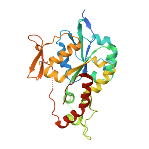
    3BD9

  • PubMed Abstract: 

    The biosynthesis of heparan sulfate (HS) involves an array of specialized sulfotransferases. Here, we present a study aimed at engineering the substrate specificity of different HS 3-O-sulfotransferase isoforms. Based on the crystal structures, we identified a pair of amino acid residues responsible for selecting the substrates. Mutations of these residues altered the substrate specificities. Our results demonstrate the feasibility of tailoring the specificity of sulfotransferases to modify HS with desired functions.


  • Organizational Affiliation

    Division of Medicinal Chemistry and Natural Products, School of Pharmacy, University of North Carolina, Chapel Hill, North Carolina 27599, USA.


Macromolecules
Find similar proteins by:  (by identity cutoff)  |  3D Structure
Entity ID: 1
MoleculeChains Sequence LengthOrganismDetailsImage
Heparan sulfate glucosamine 3-O-sulfotransferase 5280Homo sapiensMutation(s): 1 
Gene Names: HS3ST53OST5HS3OST5
EC: 2.8.2.23
UniProt & NIH Common Fund Data Resources
Find proteins for Q8IZT8 (Homo sapiens)
Explore Q8IZT8 
Go to UniProtKB:  Q8IZT8
PHAROS:  Q8IZT8
GTEx:  ENSG00000249853 
Entity Groups  
Sequence Clusters30% Identity50% Identity70% Identity90% Identity95% Identity100% Identity
UniProt GroupQ8IZT8
Sequence Annotations
Expand
  • Reference Sequence
Small Molecules
Ligands 1 Unique
IDChains Name / Formula / InChI Key2D Diagram3D Interactions
A3P
Query on A3P

Download Ideal Coordinates CCD File 
B [auth A]ADENOSINE-3'-5'-DIPHOSPHATE
C10 H15 N5 O10 P2
WHTCPDAXWFLDIH-KQYNXXCUSA-N
Experimental Data & Validation

Experimental Data

  • Method: X-RAY DIFFRACTION
  • Resolution: 2.30 Å
  • R-Value Free: 0.249 
  • R-Value Work: 0.203 
  • R-Value Observed: 0.205 
  • Space Group: P 43 21 2
Unit Cell:
Length ( Å )Angle ( ˚ )
a = 59.27α = 90
b = 59.27β = 90
c = 173.957γ = 90
Software Package:
Software NamePurpose
CNSrefinement
StructureStudiodata collection
HKL-2000data reduction
HKL-2000data scaling
MOLREPphasing

Structure Validation

View Full Validation Report


Ligand Structure Quality Assessment 


Entry History 

Deposition Data

Revision History  (Full details and data files)

  • Version 1.0: 2008-01-29
    Type: Initial release
  • Version 1.1: 2011-07-13
    Changes: Version format compliance
  • Version 1.2: 2021-10-20
    Changes: Database references, Derived calculations
  • Version 1.3: 2024-03-13
    Changes: Data collection, Source and taxonomy, Structure summary
  • Version 1.4: 2024-04-03
    Changes: Refinement description