3B8N

Structure of FepE- Bacterial Polysaccharide Co-polymerase


Experimental Data Snapshot

  • Method: X-RAY DIFFRACTION
  • Resolution: 3.10 Å
  • R-Value Free: 0.282 
  • R-Value Work: 0.254 

wwPDB Validation   3D Report Full Report


This is version 1.3 of the entry. See complete history


Literature

Bacterial polysaccharide co-polymerases share a common framework for control of polymer length

Tocilj, A.Munger, C.Proteau, A.Morona, R.Purins, L.Ajamian, E.Wagner, J.Papadopoulos, M.Van Den Bosch, L.Rubinstein, J.L.Fethiere, J.Matte, A.Cygler, M.

(2008) Nat Struct Mol Biol 15: 130-138

  • DOI: https://doi.org/10.1038/nsmb.1374
  • Primary Citation of Related Structures:  
    3B8M, 3B8N, 3B8O, 3B8P

  • PubMed Abstract: 

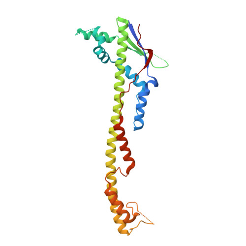
    The chain length distribution of complex polysaccharides present on the bacterial surface is determined by polysaccharide co-polymerases (PCPs) anchored in the inner membrane. We report crystal structures of the periplasmic domains of three PCPs that impart substantially different chain length distributions to surface polysaccharides. Despite very low sequence similarities, they have a common protomer structure with a long central alpha-helix extending 100 A into the periplasm. The protomers self-assemble into bell-shaped oligomers of variable sizes, with a large internal cavity. Electron microscopy shows that one of the full-length PCPs has a similar organization as that observed in the crystal for its periplasmic domain alone. Functional studies suggest that the top of the PCP oligomers is an important region for determining polysaccharide modal length. These structures provide a detailed view of components of the bacterial polysaccharide assembly machinery.


  • Organizational Affiliation

    Department of Biochemistry, McGill University, 3655 Promenade Sir William Osler, Montréal, Québec H3G 1Y6, Canada.


Macromolecules
Find similar proteins by:  (by identity cutoff)  |  3D Structure
Entity ID: 1
MoleculeChains Sequence LengthOrganismDetailsImage
Ferric enterobactin (Enterochelin) transport
A, B, C, D, E
A, B, C, D, E, F, G, H, I
279Escherichia coli O157:H7Mutation(s): 0 
Gene Names: fepE
Membrane Entity: Yes 
UniProt
Find proteins for Q8XBV8 (Escherichia coli O157:H7)
Explore Q8XBV8 
Go to UniProtKB:  Q8XBV8
Entity Groups  
Sequence Clusters30% Identity50% Identity70% Identity90% Identity95% Identity100% Identity
UniProt GroupQ8XBV8
Sequence Annotations
Expand
  • Reference Sequence
Experimental Data & Validation

Experimental Data

  • Method: X-RAY DIFFRACTION
  • Resolution: 3.10 Å
  • R-Value Free: 0.282 
  • R-Value Work: 0.254 
  • Space Group: P 32 2 1
Unit Cell:
Length ( Å )Angle ( ˚ )
a = 160.67α = 90
b = 160.67β = 90
c = 276.938γ = 120
Software Package:
Software NamePurpose
SCALEPACKdata scaling
SHARPphasing
RESOLVEphasing
REFMACrefinement
PDB_EXTRACTdata extraction
DENZOdata reduction

Structure Validation

View Full Validation Report


Entry History 

Deposition Data

Revision History  (Full details and data files)

  • Version 1.0: 2008-01-22
    Type: Initial release
  • Version 1.1: 2011-07-13
    Changes: Source and taxonomy, Version format compliance
  • Version 1.2: 2017-10-25
    Changes: Refinement description
  • Version 1.3: 2024-02-21
    Changes: Data collection, Database references, Refinement description