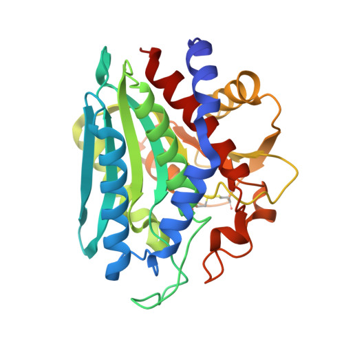
Zinc coordination geometry and ligand binding affinity: the structural and kinetic analysis of the second-shell serine 228 residue and the methionine 180 residue of the aminopeptidase from Vibrio proteolyticus.
Ataie, N.J., Hoang, Q.Q., Zahniser, M.P., Tu, Y., Milne, A., Petsko, G.A., Ringe, D.(2008) Biochemistry 47: 7673-7683
- PubMed: 18576673
- DOI: https://doi.org/10.1021/bi702188e
- Primary Citation of Related Structures:
3B35, 3B3C, 3B3S, 3B3T, 3B3V, 3B3W, 3B7I - PubMed Abstract:
The chemical properties of zinc make it an ideal metal to study the role of coordination strain in enzymatic rate enhancement. The zinc ion and the protein residues that are bound directly to the zinc ion represent a functional charge/dipole complex, and polarization of this complex, which translates to coordination distortion, may tune electrophilicity, and hence, reactivity. Conserved protein residues outside of the charge/dipole complex, such as second-shell residues, may play a role in supporting the electronic strain produced as a consequence of functional polarization. To test the correlation between charge/dipole polarity and ligand binding affinity, structure-function studies were carried out on the dizinc aminopeptidase from Vibrio proteolyticus. Alanine substitutions of S228 and M180 resulted in catalytically diminished enzymes whose crystal structures show very little change in the positions of the metal ions and the protein residues. However, more detailed inspections of the crystal structures show small positional changes that account for differences in the zinc ion coordination geometry. Measurements of the binding affinity of leucine phosphonic acid, a transition state analogue, and leucine, a product, show a correlation between coordination geometry and ligand binding affinity. These results suggest that the coordination number and polarity may tune the electrophilicity of zinc. This may have provided the evolving enzyme with the ability to discriminate between reaction coordinate species.
Organizational Affiliation:
Rosenstiel Basic Medical Sciences Research Center and Department of Biochemistry, Program in Biochemistry and Biophysics, Brandeis University, 415 South Street, Waltham, Massachusetts 02454, USA.