3AVR

Catalytic fragment of UTX/KDM6A bound with histone H3K27me3 peptide, N-oxyalylglycine, and Ni(II)


Experimental Data Snapshot

  • Method: X-RAY DIFFRACTION
  • Resolution: 1.80 Å
  • R-Value Free: 0.198 
  • R-Value Work: 0.169 
  • R-Value Observed: 0.171 

wwPDB Validation   3D Report Full Report


This is version 1.2 of the entry. See complete history


Literature

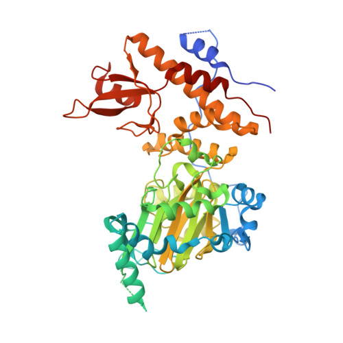
Structural basis for histone H3 Lys 27 demethylation by UTX/KDM6A

Sengoku, T.Yokoyama, S.

(2011) Genes Dev 25: 2266-2277

  • DOI: https://doi.org/10.1101/gad.172296.111
  • Primary Citation of Related Structures:  
    3AVR, 3AVS

  • PubMed Abstract: 

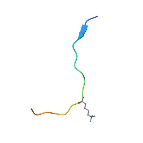
    Tri- and dimethylations of histone H3K9 (H3K9me3/2) and H3K27 (H3K27me3/2), both situated in the "A-R-Kme-S" sequence motif, mediate transcriptional repression of distinct genomic regions. H3K9me3/2 mainly governs constitutive heterochromatin formation, while H3K27me3/2 represses key developmental genes. The mechanisms by which histone-modifying enzymes selectively regulate the methylation states of H3K9 and H3K27 are poorly understood. Here we report the crystal structures of the catalytic fragment of UTX/KDM6A, an H3K27me3/2-specific demethylase, in the free and H3 peptide-bound forms. The catalytic jumonji domain binds H3 residues 25-33, recognizing H3R26, H3A29, and H3P30 in a sequence-specific manner, in addition to H3K27me3 in the catalytic pocket. A novel zinc-binding domain, conserved within the KDM6 family, binds residues 17-21 of H3. The zinc-binding domain changes its conformation upon H3 binding, and thereby recognizes the H3L20 side chain via a hydrophobic patch on its surface, which is inaccessible in the H3-free form. Mutational analyses showed that H3R17, H3L20, H3R26, H3A29, H3P30, and H3T32 are each important for demethylation. No other methyllysines in the histone tails have the same set of residues at the corresponding positions. Thus, we clarified how UTX discriminates H3K27me3/2 from the other methyllysines with distinct roles, including the near-cognate H3K9me3/2, in histones.


  • Organizational Affiliation

    RIKEN Systems and Structural Biology Center, Tsurumi, Yokohama, Japan.


Macromolecules
Find similar proteins by:  (by identity cutoff)  |  3D Structure
Entity ID: 1
MoleculeChains Sequence LengthOrganismDetailsImage
Lysine-specific demethylase 6A531Homo sapiensMutation(s): 0 
Gene Names: KDM6AUTX
EC: 1.14.11
UniProt & NIH Common Fund Data Resources
Find proteins for O15550 (Homo sapiens)
Explore O15550 
Go to UniProtKB:  O15550
PHAROS:  O15550
GTEx:  ENSG00000147050 
Entity Groups  
Sequence Clusters30% Identity50% Identity70% Identity90% Identity95% Identity100% Identity
UniProt GroupO15550
Sequence Annotations
Expand
  • Reference Sequence

Find similar proteins by:  Sequence   |   3D Structure  

Entity ID: 2
MoleculeChains Sequence LengthOrganismDetailsImage
Histone H322N/AMutation(s): 0 
UniProt & NIH Common Fund Data Resources
Find proteins for P68431 (Homo sapiens)
Explore P68431 
Go to UniProtKB:  P68431
PHAROS:  P68431
Entity Groups  
Sequence Clusters30% Identity50% Identity70% Identity90% Identity95% Identity100% Identity
UniProt GroupP68431
Sequence Annotations
Expand
  • Reference Sequence
Small Molecules
Ligands 5 Unique
IDChains Name / Formula / InChI Key2D Diagram3D Interactions
OGA
Query on OGA

Download Ideal Coordinates CCD File 
C [auth A]N-OXALYLGLYCINE
C4 H5 N O5
BIMZLRFONYSTPT-UHFFFAOYSA-N
ZN
Query on ZN

Download Ideal Coordinates CCD File 
D [auth A]ZINC ION
Zn
PTFCDOFLOPIGGS-UHFFFAOYSA-N
EDO
Query on EDO

Download Ideal Coordinates CCD File 
F [auth A]
G [auth A]
H [auth A]
I [auth A]
J [auth A]
F [auth A],
G [auth A],
H [auth A],
I [auth A],
J [auth A],
K [auth A],
L [auth A],
M [auth A],
N [auth A]
1,2-ETHANEDIOL
C2 H6 O2
LYCAIKOWRPUZTN-UHFFFAOYSA-N
NI
Query on NI

Download Ideal Coordinates CCD File 
E [auth A]NICKEL (II) ION
Ni
VEQPNABPJHWNSG-UHFFFAOYSA-N
CL
Query on CL

Download Ideal Coordinates CCD File 
O [auth A],
P [auth A]
CHLORIDE ION
Cl
VEXZGXHMUGYJMC-UHFFFAOYSA-M
Modified Residues  1 Unique
IDChains TypeFormula2D DiagramParent
M3L
Query on M3L
B
L-PEPTIDE LINKINGC9 H21 N2 O2LYS
Experimental Data & Validation

Experimental Data

  • Method: X-RAY DIFFRACTION
  • Resolution: 1.80 Å
  • R-Value Free: 0.198 
  • R-Value Work: 0.169 
  • R-Value Observed: 0.171 
  • Space Group: P 21 21 21
Unit Cell:
Length ( Å )Angle ( ˚ )
a = 78.905α = 90
b = 83.081β = 90
c = 95.057γ = 90
Software Package:
Software NamePurpose
HKL-2000data collection
PHASERphasing
PHENIXrefinement
HKL-2000data reduction
HKL-2000data scaling

Structure Validation

View Full Validation Report


Entry History 

Deposition Data

Revision History  (Full details and data files)

  • Version 1.0: 2011-10-19
    Type: Initial release
  • Version 1.1: 2011-12-14
    Changes: Database references
  • Version 1.2: 2023-11-01
    Changes: Data collection, Database references, Derived calculations, Refinement description