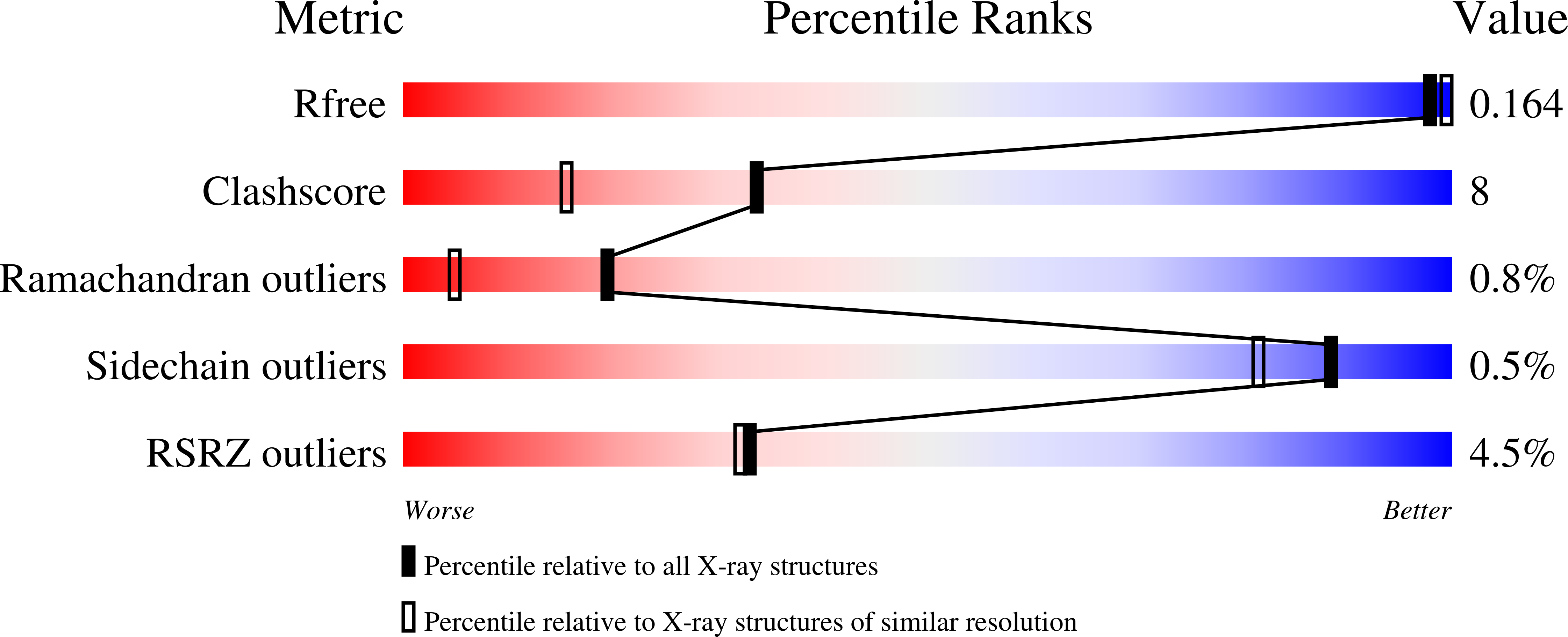
3ATG

endo-1,3-beta-glucanase from Cellulosimicrobium cellulans


Experimental Data Snapshot

  • Method: X-RAY DIFFRACTION
  • Resolution: 1.66 Å
  • R-Value Free: 0.166 
  • R-Value Work: 0.145 
  • R-Value Observed: 0.147 

wwPDB Validation   3D Report Full Report


This is version 1.2 of the entry. See complete history


Literature

Structural and thermodynamic characterization of endo-1,3-beta-glucanase: Insights into the substrate recognition mechanism.

Oda, M.Inaba, S.Kamiya, N.Bekker, G.J.Mikami, B.

(2018) Biochim Biophys Acta 1866: 415-425

  • DOI: https://doi.org/10.1016/j.bbapap.2017.12.004
  • Primary Citation of Related Structures:  
    3ATG

  • PubMed Abstract: 

    Endo-1,3-β-glucanase from Cellulosimicrobium cellulans is composed of a catalytic domain and a carbohydrate-binding module. We have determined the X-ray crystal structure of the catalytic domain at a high resolution of 1.66Å. The overall fold is a sandwich-like β-jelly roll architecture like the enzymes in the glycoside hydrolase family 16. The substrate-binding cleft has a length and a width of ~28 and ~15Å, respectively, which is thought to be capable of accommodating at least six glucopyranose units. Laminarihexaose was placed into the substrate-binding cleft, namely at the subsites +2 to -4 from the reducing end, and the complex structure was analyzed using molecular dynamics simulations (MD) and using a rotamer search of the pocket. During the MD simulations, the substrate fluctuated more than the enzyme, where the residues at the subsites toward the non-reducing end fluctuated more than those toward the reducing end. Little conformational change of the protein was observed for the subsites +1 and +2, indicating that the glucose's position could be tightly restricted inside the pocket. Substrate binding experiments using isothermal titration calorimetry showed that the binding affinity of laminaritriose was higher than that of laminaribiose and similar to those of other longer laminarioligosaccharides. Taken together, the substrates mainly bind to the subsites -1 to -3 with the highest affinity, while the part bound to the reducing end would be hydrolyzed.


  • Organizational Affiliation

    Graduate School of Life and Environmental Sciences, Kyoto Prefectural University, 1-5 Hangi-cho, Shimogamo, Sakyo, Kyoto, Kyoto 606-8522, Japan. Electronic address: oda@kpu.ac.jp.


Macromolecules
Find similar proteins by:  (by identity cutoff)  |  3D Structure
Entity ID: 1
MoleculeChains Sequence LengthOrganismDetailsImage
GLUCANASE256Cellulosimicrobium cellulansMutation(s): 0 
EC: 3.2.1.39
UniProt
Find proteins for G1ED17 (Cellulosimicrobium cellulans)
Explore G1ED17 
Go to UniProtKB:  G1ED17
Entity Groups  
Sequence Clusters30% Identity50% Identity70% Identity90% Identity95% Identity100% Identity
UniProt GroupG1ED17
Sequence Annotations
Expand
  • Reference Sequence
Small Molecules
Ligands 3 Unique
IDChains Name / Formula / InChI Key2D Diagram3D Interactions
PO4
Query on PO4

Download Ideal Coordinates CCD File 
B [auth A],
C [auth A],
D [auth A],
E [auth A],
F [auth A]
PHOSPHATE ION
O4 P
NBIIXXVUZAFLBC-UHFFFAOYSA-K
GOL
Query on GOL

Download Ideal Coordinates CCD File 
G [auth A],
H [auth A],
I [auth A]
GLYCEROL
C3 H8 O3
PEDCQBHIVMGVHV-UHFFFAOYSA-N
CA
Query on CA

Download Ideal Coordinates CCD File 
J [auth A]CALCIUM ION
Ca
BHPQYMZQTOCNFJ-UHFFFAOYSA-N
Experimental Data & Validation

Experimental Data

  • Method: X-RAY DIFFRACTION
  • Resolution: 1.66 Å
  • R-Value Free: 0.166 
  • R-Value Work: 0.145 
  • R-Value Observed: 0.147 
  • Space Group: P 43
Unit Cell:
Length ( Å )Angle ( ˚ )
a = 71.309α = 90
b = 71.309β = 90
c = 60.069γ = 90
Software Package:
Software NamePurpose
HKL-2000data collection
SOLVEphasing
PHENIXrefinement
HKL-2000data reduction
HKL-2000data scaling

Structure Validation

View Full Validation Report


Entry History 

Deposition Data

Revision History  (Full details and data files)

  • Version 1.0: 2012-01-18
    Type: Initial release
  • Version 1.1: 2018-01-03
    Changes: Database references
  • Version 1.2: 2024-03-13
    Changes: Data collection, Database references, Derived calculations