3ALO

Crystal structure of human non-phosphorylated MKK4 kinase domain ternary complex with AMP-PNP and p38 peptide


Experimental Data Snapshot

  • Method: X-RAY DIFFRACTION
  • Resolution: 2.60 Å
  • R-Value Free: 0.271 
  • R-Value Work: 0.206 
  • R-Value Observed: 0.209 

wwPDB Validation   3D Report Full Report


Ligand Structure Quality Assessment 


This is version 1.3 of the entry. See complete history


Literature

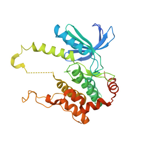
Crystal structures of MKK4 kinase domain reveal that substrate peptide binds to an allosteric site and induces an auto-inhibition state

Matsumoto, T.Kinoshita, T.Kirii, Y.Yokota, K.Hamada, K.Tada, T.

(2010) Biochem Biophys Res Commun 400: 369-373

  • DOI: https://doi.org/10.1016/j.bbrc.2010.08.071
  • Primary Citation of Related Structures:  
    3ALN, 3ALO

  • PubMed Abstract: 

    MKK4 activates both JNKs and p38s. We determined the crystal structures of human non-phosphorylated MKK4 kinase domain (npMKK4) complexed with AMP-PNP (npMKK4/AMP) and a ternary complex of npMKK4, AMP-PNP and p38α peptide (npMKK4/AMP/p38). These crystal structures revealed that the p38α peptide-bound npMKK4 at the allosteric site rather than at the putative substrate binding site and induced an auto-inhibition state. While the activation loop of the npMKK4/AMP complex was disordered, in the npMKK4/AMP/p38 complex it configured a long α-helix, which prevented substrate access to the active site and αC-helix movement to the active configuration of MKK4.


  • Organizational Affiliation

    PharmAxess, Inc., 3-9-12 Matsubara-cho, Akishima, Tokyo 196-8666, Japan.


Macromolecules
Find similar proteins by:  (by identity cutoff)  |  3D Structure
Entity ID: 1
MoleculeChains Sequence LengthOrganismDetailsImage
Dual specificity mitogen-activated protein kinase kinase 4327Homo sapiensMutation(s): 0 
Gene Names: JNKK1MAP2K4MEK4MKK4PRKMK4SERK1
EC: 2.7.12.2
UniProt & NIH Common Fund Data Resources
Find proteins for P45985 (Homo sapiens)
Explore P45985 
Go to UniProtKB:  P45985
PHAROS:  P45985
GTEx:  ENSG00000065559 
Entity Groups  
Sequence Clusters30% Identity50% Identity70% Identity90% Identity95% Identity100% Identity
UniProt GroupP45985
Sequence Annotations
Expand
  • Reference Sequence

Find similar proteins by:  Sequence   |   3D Structure  

Entity ID: 2
MoleculeChains Sequence LengthOrganismDetailsImage
p38 peptideB [auth E]8Homo sapiensMutation(s): 0 
Sequence Annotations
Expand
  • Reference Sequence
Small Molecules
Ligands 2 Unique
IDChains Name / Formula / InChI Key2D Diagram3D Interactions
ANP
Query on ANP

Download Ideal Coordinates CCD File 
D [auth A]PHOSPHOAMINOPHOSPHONIC ACID-ADENYLATE ESTER
C10 H17 N6 O12 P3
PVKSNHVPLWYQGJ-KQYNXXCUSA-N
MG
Query on MG

Download Ideal Coordinates CCD File 
C [auth A]MAGNESIUM ION
Mg
JLVVSXFLKOJNIY-UHFFFAOYSA-N
Modified Residues  1 Unique
IDChains TypeFormula2D DiagramParent
TPO
Query on TPO
B [auth E]L-PEPTIDE LINKINGC4 H10 N O6 PTHR
Experimental Data & Validation

Experimental Data

  • Method: X-RAY DIFFRACTION
  • Resolution: 2.60 Å
  • R-Value Free: 0.271 
  • R-Value Work: 0.206 
  • R-Value Observed: 0.209 
  • Space Group: C 2 2 21
Unit Cell:
Length ( Å )Angle ( ˚ )
a = 68.776α = 90
b = 117.985β = 90
c = 81.793γ = 90
Software Package:
Software NamePurpose
MOLREPphasing
REFMACrefinement
CrystalCleardata reduction
CrystalCleardata scaling

Structure Validation

View Full Validation Report


Ligand Structure Quality Assessment 


Entry History 

Deposition Data

Revision History  (Full details and data files)

  • Version 1.0: 2010-10-27
    Type: Initial release
  • Version 1.1: 2011-07-13
    Changes: Version format compliance
  • Version 1.2: 2017-10-11
    Changes: Refinement description
  • Version 1.3: 2023-11-01
    Changes: Data collection, Database references, Derived calculations, Refinement description