3AJN

Structural basis of glycine amide on suppression of protein aggregation by high resolution X-ray analysis


Experimental Data Snapshot

  • Method: X-RAY DIFFRACTION
  • Resolution: 1.05 Å
  • R-Value Free: 0.163 
  • R-Value Work: 0.146 
  • R-Value Observed: 0.147 

wwPDB Validation   3D Report Full Report


This is version 1.2 of the entry. See complete history


Literature

Glycine amide shielding on the aromatic surfaces of lysozyme: Implication for suppression of protein aggregation

Ito, L.Shiraki, K.Makino, M.Hasegawa, K.Kumasaka, T.

(2011) FEBS Lett 585: 555-560

  • DOI: https://doi.org/10.1016/j.febslet.2011.01.008
  • Primary Citation of Related Structures:  
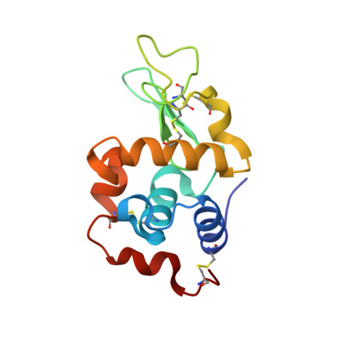
    3AJN

  • PubMed Abstract: 

    Glycine amide (GlyAd), a typically amidated amino acid, is a versatile additive that suppresses protein aggregation during refolding, heat treatment, and crystallization. In spite of its effectiveness, the exact mechanism by which GlyAd suppresses protein aggregation remains to be elucidated. Here, we show the crystal structure of the GlyAd-lysozyme complex by high resolution X-ray crystallographic analysis at a 1.05Å resolution. GlyAd bound to the lysozyme surface near aromatic residues and decreased the amount of bound waters and increased the mobility of protein. Arg and GlyAd molecules are different in binding sites and patterns from glycerol and related compounds, indicating that decreasing hydrophobic patches might be involved in suppression of protein aggregation.


  • Organizational Affiliation

    Japan Synchrotron Radiation Research Institute (SPring-8), Sayo, Hyogo, Japan. l-ito@spring8.or.jp


Macromolecules
Find similar proteins by:  (by identity cutoff)  |  3D Structure
Entity ID: 1
MoleculeChains Sequence LengthOrganismDetailsImage
Lysozyme C129Gallus gallusMutation(s): 0 
EC: 3.2.1.17
UniProt
Find proteins for P00698 (Gallus gallus)
Explore P00698 
Go to UniProtKB:  P00698
Entity Groups  
Sequence Clusters30% Identity50% Identity70% Identity90% Identity95% Identity100% Identity
UniProt GroupP00698
Sequence Annotations
Expand
  • Reference Sequence
Experimental Data & Validation

Experimental Data

  • Method: X-RAY DIFFRACTION
  • Resolution: 1.05 Å
  • R-Value Free: 0.163 
  • R-Value Work: 0.146 
  • R-Value Observed: 0.147 
  • Space Group: P 43 21 2
Unit Cell:
Length ( Å )Angle ( ˚ )
a = 78.51α = 90
b = 78.51β = 90
c = 36.962γ = 90
Software Package:
Software NamePurpose
HKL-2000data collection
MOLREPphasing
REFMACrefinement
HKL-2000data reduction
HKL-2000data scaling

Structure Validation

View Full Validation Report


Entry History 

Deposition Data

Revision History  (Full details and data files)

  • Version 1.0: 2011-02-16
    Type: Initial release
  • Version 1.1: 2011-07-13
    Changes: Version format compliance
  • Version 1.2: 2023-11-01
    Changes: Data collection, Database references, Derived calculations, Refinement description