3AI4

Crystal structure of yeast enhanced green fluorescent protein - mouse polymerase iota ubiquitin binding motif fusion protein


Experimental Data Snapshot

  • Method: X-RAY DIFFRACTION
  • Resolution: 1.60 Å
  • R-Value Free: 0.221 
  • R-Value Work: 0.189 
  • R-Value Observed: 0.191 

wwPDB Validation   3D Report Full Report


This is version 1.4 of the entry. See complete history


Literature

Crystallization of small proteins assisted by green fluorescent protein

Suzuki, N.Hiraki, M.Yamada, Y.Matsugaki, N.Igarashi, N.Kato, R.Dikic, I.Drew, D.Iwata, S.Wakatsuki, S.Kawasaki, M.

(2010) Acta Crystallogr D Biol Crystallogr 66: 1059-1066

  • DOI: https://doi.org/10.1107/S0907444910032944
  • Primary Citation of Related Structures:  
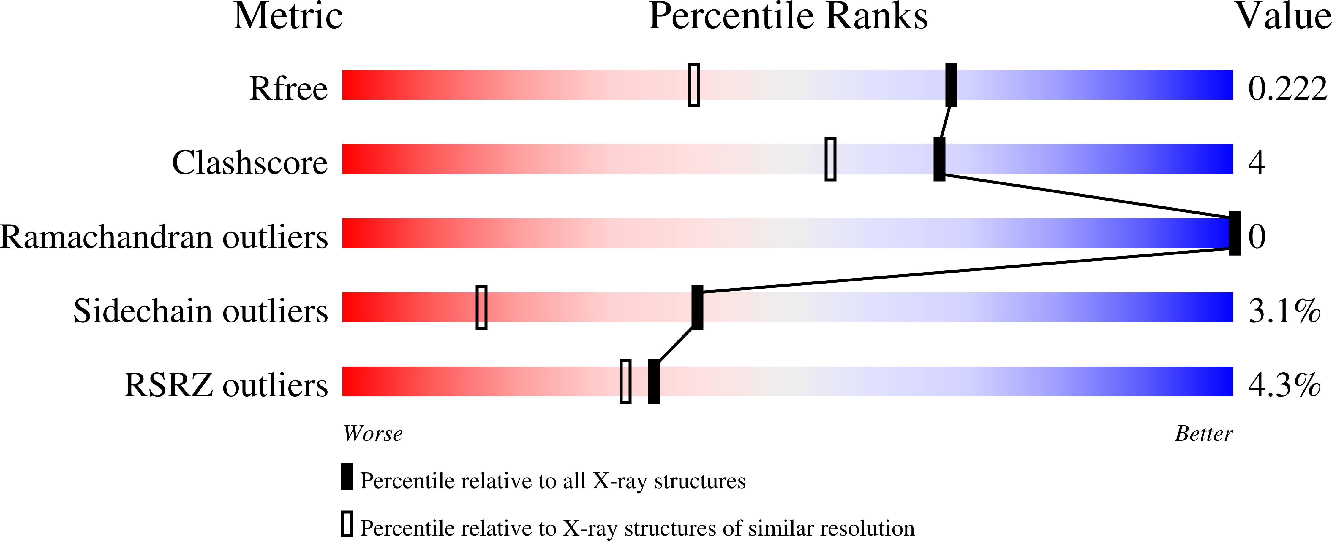
    3AI4, 3AI5

  • PubMed Abstract: 

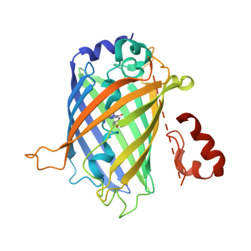
    The generation of crystal lattice contacts by proteinaceous tags fused to target proteins is an attractive approach to aid in the crystallization of otherwise intractable proteins. Here, the use of green fluorescent protein (GFP) fusions for this purpose is demonstrated, using ubiquitin and the ubiquitin-binding motif (UBM) of Y-family polymerase ι as examples. The structure of the GFP-ubiquitin fusion protein revealed that the crystal lattice was formed by GFP moieties. Ubiquitin was accommodated in the lattice through interactions with the peripheral loops of GFP. However, in the GFP-UBM fusion crystal UBM formed extensive interactions with GFP and these interactions, together with UBM dimerization, mediated the crystal packing. Interestingly, the tyrosine residues that are involved in mediating crystal contacts in both GFP-ubiquitin and GFP-UBM crystals are arranged in a belt on the surface of the β-barrel structure of GFP. Therefore, it is likely that GFP can assist in the crystallization of small proteins and of protein domains in general.


  • Organizational Affiliation

    Structural Biology Research Center, Photon Factory, Institute of Materials Structure Science, High Energy Accelerator Research Organization (KEK), Tsukuba, Ibaraki 305-0801, Japan.


Macromolecules
Find similar proteins by:  (by identity cutoff)  |  3D Structure
Entity ID: 1
MoleculeChains Sequence LengthOrganismDetailsImage
yeast enhanced green fluorescent protein,DNA polymerase iota283Aequorea victoriaMus musculus
This entity is chimeric
Mutation(s): 0 
UniProt & NIH Common Fund Data Resources
Find proteins for P42212 (Aequorea victoria)
Explore P42212 
Go to UniProtKB:  P42212
Find proteins for Q6R3M4 (Mus musculus)
Explore Q6R3M4 
Go to UniProtKB:  Q6R3M4
IMPC:  MGI:1347081
Entity Groups  
Sequence Clusters30% Identity50% Identity70% Identity90% Identity95% Identity100% Identity
UniProt GroupsP42212Q6R3M4
Sequence Annotations
Expand
  • Reference Sequence
Small Molecules
Ligands 1 Unique
IDChains Name / Formula / InChI Key2D Diagram3D Interactions
SO4
Query on SO4

Download Ideal Coordinates CCD File 
B [auth A]SULFATE ION
O4 S
QAOWNCQODCNURD-UHFFFAOYSA-L
Modified Residues  1 Unique
IDChains TypeFormula2D DiagramParent
CR2
Query on CR2
A
L-PEPTIDE LINKINGC13 H13 N3 O4GLY, TYR, GLY
Experimental Data & Validation

Experimental Data

  • Method: X-RAY DIFFRACTION
  • Resolution: 1.60 Å
  • R-Value Free: 0.221 
  • R-Value Work: 0.189 
  • R-Value Observed: 0.191 
  • Space Group: P 42 21 2
Unit Cell:
Length ( Å )Angle ( ˚ )
a = 87.457α = 90
b = 87.457β = 90
c = 83.611γ = 90
Software Package:
Software NamePurpose
ADSCdata collection
MOLREPphasing
REFMACrefinement
HKL-2000data reduction
HKL-2000data scaling

Structure Validation

View Full Validation Report


Entry History 

Deposition Data

Revision History  (Full details and data files)

  • Version 1.0: 2010-09-29
    Type: Initial release
  • Version 1.1: 2011-07-13
    Changes: Version format compliance
  • Version 1.2: 2017-06-14
    Changes: Database references, Source and taxonomy, Structure summary
  • Version 1.3: 2023-11-01
    Changes: Data collection, Database references, Derived calculations, Refinement description
  • Version 1.4: 2023-11-15
    Changes: Data collection