


Crystal structure analysis of the fluorescent protein KillerRed
Sakai, N., Takemoto, K., Matsuda, T., Kitago, Y., Ayabe, T., Nagai, T.To be published.
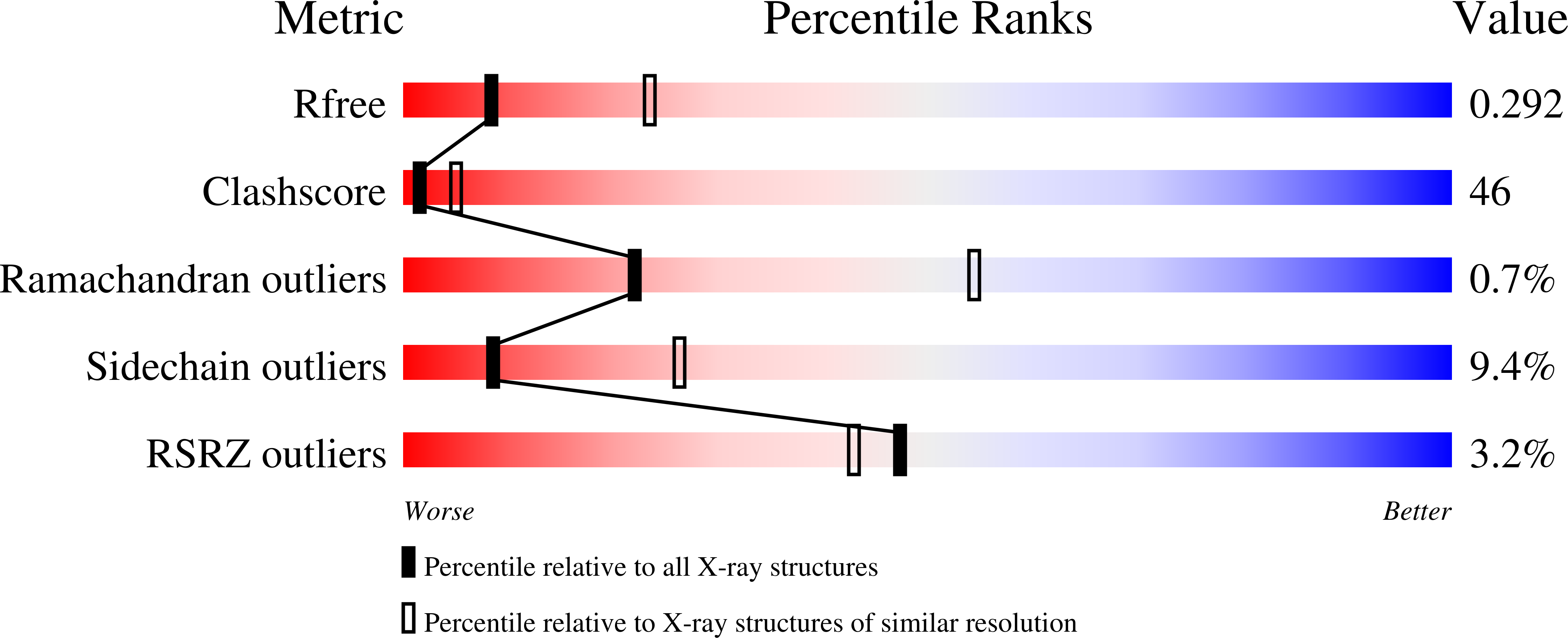
Experimental Data Snapshot
wwPDB Validation 3D Report Full Report
Entity ID: 1 | |||||
|---|---|---|---|---|---|
| Molecule | Chains | Sequence Length | Organism | Details | Image |
| KillerRed | 238 | Anthomedusae sp. DC-2005 | Mutation(s): 1 | 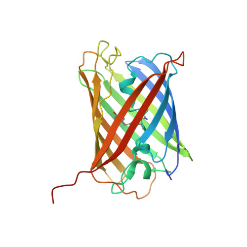 | |
UniProt | |||||
Find proteins for Q2TCH5 (Anthomedusae sp. DC-2005) Explore Q2TCH5 Go to UniProtKB: Q2TCH5 | |||||
Entity Groups | |||||
| Sequence Clusters | 30% Identity50% Identity70% Identity90% Identity95% Identity100% Identity | ||||
| UniProt Group | Q2TCH5 | ||||
Sequence AnnotationsExpand | |||||
| |||||
| Modified Residues 1 Unique | |||||
|---|---|---|---|---|---|
| ID | Chains | Type | Formula | 2D Diagram | Parent |
| CRQ Query on CRQ | A, B | L-PEPTIDE LINKING | C16 H16 N4 O5 |  | GLN, TYR, GLY |
| Length ( Å ) | Angle ( ˚ ) |
|---|---|
| a = 123.464 | α = 90 |
| b = 123.464 | β = 90 |
| c = 110.171 | γ = 120 |
| Software Name | Purpose |
|---|---|
| CrystalClear | data collection |
| SHELXDE | phasing |
| REFMAC | refinement |
| HKL-2000 | data reduction |
| SCALEPACK | data scaling |
RCSB PDB (citation) is hosted by
RCSB PDB is a member of the

