


In-Crystal Chemical Ligation for Drug Discovery
Yamane, J., Ooyabu, N., Yao, M., Takemoto, H., Tanaka, I.To be published.
Experimental Data Snapshot
Entity ID: 1 | |||||
|---|---|---|---|---|---|
| Molecule | Chains | Sequence Length | Organism | Details | Image |
| Cationic trypsin | 223 | Bos taurus | Mutation(s): 0 EC: 3.4.21.4 | 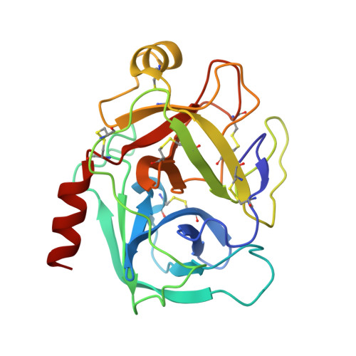 | |
UniProt | |||||
Find proteins for P00760 (Bos taurus) Explore P00760 Go to UniProtKB: P00760 | |||||
Entity Groups | |||||
| Sequence Clusters | 30% Identity50% Identity70% Identity90% Identity95% Identity100% Identity | ||||
| UniProt Group | P00760 | ||||
Sequence AnnotationsExpand | |||||
| |||||
| Ligands 4 Unique | |||||
|---|---|---|---|---|---|
| ID | Chains | Name / Formula / InChI Key | 2D Diagram | 3D Interactions | |
| O04 Query on O04 | E [auth A] | (E)-4-((tetrahydro-2H-pyran-2-yloxyimino)methyl)benzimidamide C13 H17 N3 O2 WEPKQSGLGGDMSU-RGZVIEDOSA-N |  | ||
| SO4 Query on SO4 | D [auth A] | SULFATE ION O4 S QAOWNCQODCNURD-UHFFFAOYSA-L |  | ||
| GOL Query on GOL | C [auth A] | GLYCEROL C3 H8 O3 PEDCQBHIVMGVHV-UHFFFAOYSA-N |  | ||
| CA Query on CA | B [auth A] | CALCIUM ION Ca BHPQYMZQTOCNFJ-UHFFFAOYSA-N |  | ||
| Length ( Å ) | Angle ( ˚ ) |
|---|---|
| a = 54.447 | α = 90 |
| b = 58.542 | β = 90 |
| c = 66.562 | γ = 90 |
| Software Name | Purpose |
|---|---|
| LAFIRE | model building |
| REFMAC | refinement |
| HKL-2000 | data reduction |
| HKL-2000 | data scaling |
| LAFIRE | phasing |
RCSB PDB (citation) is hosted by
RCSB PDB is a member of the

