3A7J

Human MST3 kinase in complex with MnADP


Experimental Data Snapshot

  • Method: X-RAY DIFFRACTION
  • Resolution: 1.50 Å
  • R-Value Free: 0.210 
  • R-Value Work: 0.186 
  • R-Value Observed: 0.191 

wwPDB Validation   3D Report Full Report


This is version 1.3 of the entry. See complete history


Literature

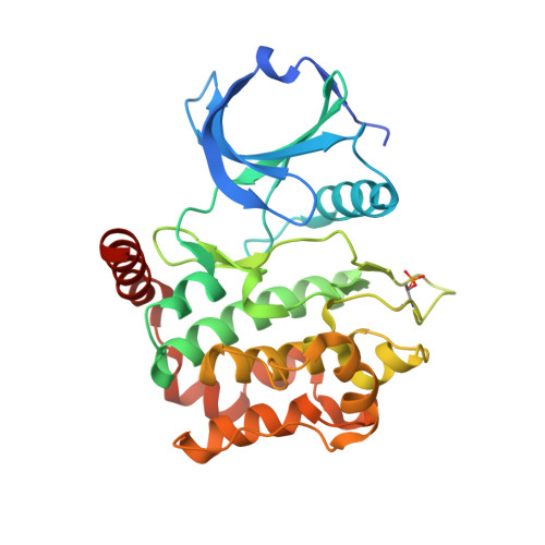
Structures of human MST3 kinase in complex with adenine, ADP and Mn2+.

Ko, T.P.Jeng, W.Y.Liu, C.I.Lai, M.D.Wu, C.L.Chang, W.J.Shr, H.L.Lu, T.J.Wang, A.H.J.

(2010) Acta Crystallogr D Biol Crystallogr 66: 145-154

  • DOI: https://doi.org/10.1107/S0907444909047507
  • Primary Citation of Related Structures:  
    3A7F, 3A7G, 3A7H, 3A7I, 3A7J

  • PubMed Abstract: 

    The MST family is a subclass of mammalian serine/threonine kinases that are related to the yeast sterile-20 protein and are implicated in regulating cell growth and transformation. The MST3 protein contains a 300-residue catalytic domain and a 130-residue regulatory domain, which can be cleaved by caspase and activated by autophosphorylation, promoting apoptosis. Here, five crystal structures of the catalytic domain of MST3 are presented, including a complex with ADP and manganese, a unique cofactor preferred by the enzyme, and a complex with adenine. Similar to other protein kinases, the catalytic domain of MST3 folds into two lobes: the smaller N lobe forms the nucleotide-binding site and the larger C lobe recognizes the polypeptide substrate. The bound ADP and Mn(2+) ions are covered by a glycine-rich loop and held in place by Asn149 and Asp162. A different orientation was observed for the ligand in the MST3-adenine complex. In the activation loop, the side chain of Thr178 is phosphorylated and is sandwiched by Arg143 and Arg176. Comparison of this structure with other similar kinase structures shows a 180 degrees rotation of the loop, leading to activation of the enzyme. The well defined protein-ligand interactions also provide useful information for the design of potent inhibitors.


  • Organizational Affiliation

    Institute of Biological Chemistry, Academia Sinica, Taipei 115, Taiwan.


Macromolecules
Find similar proteins by:  (by identity cutoff)  |  3D Structure
Entity ID: 1
MoleculeChains Sequence LengthOrganismDetailsImage
Serine/threonine kinase 24 (STE20 homolog, yeast)303Homo sapiensMutation(s): 0 
Gene Names: STK24
UniProt & NIH Common Fund Data Resources
Find proteins for Q9Y6E0 (Homo sapiens)
Explore Q9Y6E0 
Go to UniProtKB:  Q9Y6E0
PHAROS:  Q9Y6E0
GTEx:  ENSG00000102572 
Entity Groups  
Sequence Clusters30% Identity50% Identity70% Identity90% Identity95% Identity100% Identity
UniProt GroupQ9Y6E0
Sequence Annotations
Expand
  • Reference Sequence
Small Molecules
Modified Residues  1 Unique
IDChains TypeFormula2D DiagramParent
TPO
Query on TPO
A
L-PEPTIDE LINKINGC4 H10 N O6 PTHR
Experimental Data & Validation

Experimental Data

  • Method: X-RAY DIFFRACTION
  • Resolution: 1.50 Å
  • R-Value Free: 0.210 
  • R-Value Work: 0.186 
  • R-Value Observed: 0.191 
  • Space Group: P 1 21 1
Unit Cell:
Length ( Å )Angle ( ˚ )
a = 47.816α = 90
b = 53.527β = 109.95
c = 60.634γ = 90
Software Package:
Software NamePurpose
HKL-2000data collection
CNSrefinement
HKL-2000data reduction
HKL-2000data scaling
CNSphasing

Structure Validation

View Full Validation Report


Entry History 

Deposition Data

Revision History  (Full details and data files)

  • Version 1.0: 2010-02-02
    Type: Initial release
  • Version 1.1: 2011-07-13
    Changes: Version format compliance
  • Version 1.2: 2012-04-04
    Changes: Database references
  • Version 1.3: 2023-11-01
    Changes: Data collection, Database references, Derived calculations, Refinement description