3A61

Crystal structure of unphosphorylated p70S6K1 (Form II)


Experimental Data Snapshot

  • Method: X-RAY DIFFRACTION
  • Resolution: 3.43 Å
  • R-Value Free: 0.349 
  • R-Value Work: 0.265 
  • R-Value Observed: 0.274 

wwPDB Validation   3D Report Full Report


Ligand Structure Quality Assessment 


This is version 1.2 of the entry. See complete history


Literature

Structural basis of human p70 ribosomal S6 kinase-1 regulation by activation loop phosphorylation.

Sunami, T.Byrne, N.Diehl, R.E.Funabashi, K.Hall, D.L.Ikuta, M.Patel, S.B.Shipman, J.M.Smith, R.F.Takahashi, I.Zugay-Murphy, J.Iwasawa, Y.Lumb, K.J.Munshi, S.K.Sharma, S.

(2010) J Biol Chem 285: 4587-4594

  • DOI: https://doi.org/10.1074/jbc.M109.040667
  • Primary Citation of Related Structures:  
    3A60, 3A61, 3A62

  • PubMed Abstract: 

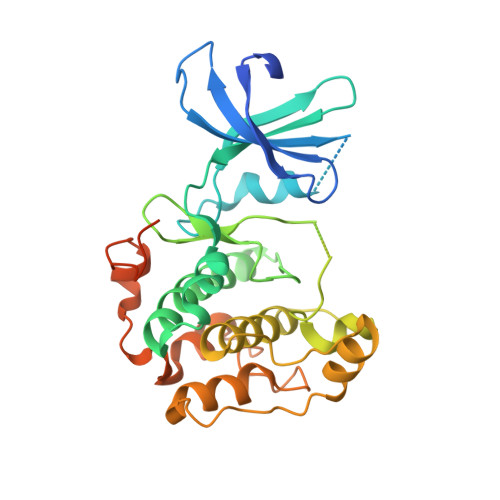
    p70 ribosomal S6 kinase (p70S6K) is a downstream effector of the mTOR signaling pathway involved in cell proliferation, cell growth, cell-cycle progression, and glucose homeostasis. Multiple phosphorylation events within the catalytic, autoinhibitory, and hydrophobic motif domains contribute to the regulation of p70S6K. We report the crystal structures of the kinase domain of p70S6K1 bound to staurosporine in both the unphosphorylated state and in the 3'-phosphoinositide-dependent kinase-1-phosphorylated state in which Thr-252 of the activation loop is phosphorylated. Unphosphorylated p70S6K1 exists in two crystal forms, one in which the p70S6K1 kinase domain exists as a monomer and the other as a domain-swapped dimer. The crystal structure of the partially activated kinase domain that is phosphorylated within the activation loop reveals conformational ordering of the activation loop that is consistent with a role in activation. The structures offer insights into the structural basis of the 3'-phosphoinositide-dependent kinase-1-induced activation of p70S6K and provide a platform for the rational structure-guided design of specific p70S6K inhibitors.


  • Organizational Affiliation

    Department of Chemistry, Tsukuba Research Institute, Banyu Pharmaceutical Company, Limited, Tsukuba, Ibaraki, 300-2611, Japan. sunami.tomoko@jaea.go.jp


Macromolecules
Find similar proteins by:  (by identity cutoff)  |  3D Structure
Entity ID: 1
MoleculeChains Sequence LengthOrganismDetailsImage
Ribosomal protein S6 kinase beta-1327Homo sapiensMutation(s): 0 
EC: 2.7.11.1
UniProt & NIH Common Fund Data Resources
Find proteins for P23443 (Homo sapiens)
Explore P23443 
Go to UniProtKB:  P23443
PHAROS:  P23443
GTEx:  ENSG00000108443 
Entity Groups  
Sequence Clusters30% Identity50% Identity70% Identity90% Identity95% Identity100% Identity
UniProt GroupP23443
Sequence Annotations
Expand
  • Reference Sequence
Small Molecules
Ligands 1 Unique
IDChains Name / Formula / InChI Key2D Diagram3D Interactions
STU
Query on STU

Download Ideal Coordinates CCD File 
B [auth A]STAUROSPORINE
C28 H26 N4 O3
HKSZLNNOFSGOKW-FYTWVXJKSA-N
Experimental Data & Validation

Experimental Data

  • Method: X-RAY DIFFRACTION
  • Resolution: 3.43 Å
  • R-Value Free: 0.349 
  • R-Value Work: 0.265 
  • R-Value Observed: 0.274 
  • Space Group: P 21 21 21
Unit Cell:
Length ( Å )Angle ( ˚ )
a = 56.636α = 90
b = 63.014β = 90
c = 98.349γ = 90
Software Package:
Software NamePurpose
CrystalCleardata collection
AMoREphasing
CNXrefinement
HKL-2000data reduction
HKL-2000data scaling

Structure Validation

View Full Validation Report


Ligand Structure Quality Assessment 


Entry History 

Revision History  (Full details and data files)

  • Version 1.0: 2009-10-27
    Type: Initial release
  • Version 1.1: 2011-07-13
    Changes: Version format compliance
  • Version 1.2: 2023-11-01
    Changes: Data collection, Database references, Derived calculations, Refinement description