3A58

Crystal structure of Sec3p - Rho1p complex from Saccharomyces cerevisiae


Experimental Data Snapshot

  • Method: X-RAY DIFFRACTION
  • Resolution: 2.60 Å
  • R-Value Free: 0.265 
  • R-Value Work: 0.234 
  • R-Value Observed: 0.235 

wwPDB Validation   3D Report Full Report


Ligand Structure Quality Assessment 


This is version 1.3 of the entry. See complete history


Literature

Structural basis for the Rho- and phosphoinositide-dependent localization of the exocyst subunit Sec3

Yamashita, M.Kurokawa, K.Sato, Y.Yamagata, A.Mimura, H.Yoshikawa, A.Sato, K.Nakano, A.Fukai, S.

(2010) Nat Struct Mol Biol 17: 180-186

  • DOI: https://doi.org/10.1038/nsmb.1722
  • Primary Citation of Related Structures:  
    3A58

  • PubMed Abstract: 

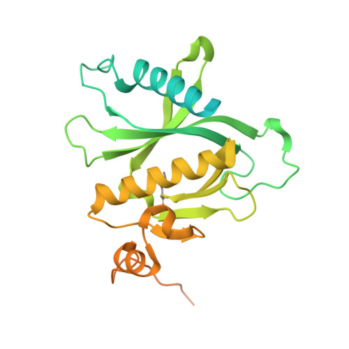
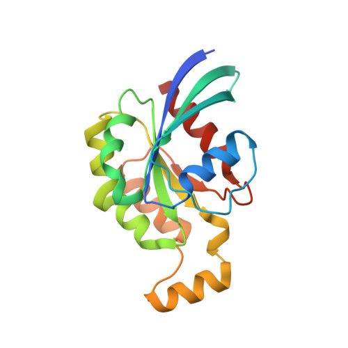
    The exocyst complex is a hetero-octameric protein complex that functions during cell polarization by tethering the secretory vesicle to the target membrane. The yeast exocyst subunit Sec3 binds to phosphatidylinositol 4,5-bisphosphate (PtdIns(4,5)P(2)) and the small GTPases Rho1 and Cdc42 via its N-terminal domain (Sec3-N), and these interactions target Sec3 to the plasma membrane. Here we report the crystal structure of the Sec3-N in complex with Rho1 at 2.6-A resolution. Sec3-N adopts a pleckstrin homology (PH) fold, despite having no detectable sequence homology with other PH domains of known structure. Clusters of conserved basic residues constitute a positively charged cleft, which was identified as a binding site for PtdIns(4,5)P(2). Residues Phe77, Ile115 and Leu131 of Sec3 bind to an extended hydrophobic surface formed around switch regions I and II of Rho1. To our knowledge, these are the first structural insights into how an exocyst subunit might interact with both protein and phospholipid factors on the target membrane.


  • Organizational Affiliation

    Structural Biology Laboratory, Life Science Division, Synchrotron Radiation Research Organization and Institute of Molecular and Cellular Biosciences, The University of Tokyo, Tokyo, Japan.


Macromolecules
Find similar proteins by:  (by identity cutoff)  |  3D Structure
Entity ID: 1
MoleculeChains Sequence LengthOrganismDetailsImage
Exocyst complex component SEC3
A, C, E
320Saccharomyces cerevisiaeMutation(s): 0 
Gene Names: SEC3PSL1YER008C
UniProt
Find proteins for P33332 (Saccharomyces cerevisiae (strain ATCC 204508 / S288c))
Explore P33332 
Go to UniProtKB:  P33332
Entity Groups  
Sequence Clusters30% Identity50% Identity70% Identity90% Identity95% Identity100% Identity
UniProt GroupP33332
Sequence Annotations
Expand
  • Reference Sequence
Find similar proteins by:  (by identity cutoff)  |  3D Structure
Entity ID: 2
MoleculeChains Sequence LengthOrganismDetailsImage
GTP-binding protein RHO1
B, D, F
188Saccharomyces cerevisiaeMutation(s): 1 
Gene Names: RHO1YPR165WP9325.3
UniProt
Find proteins for P06780 (Saccharomyces cerevisiae (strain ATCC 204508 / S288c))
Explore P06780 
Go to UniProtKB:  P06780
Entity Groups  
Sequence Clusters30% Identity50% Identity70% Identity90% Identity95% Identity100% Identity
UniProt GroupP06780
Sequence Annotations
Expand
  • Reference Sequence
Small Molecules
Ligands 3 Unique
IDChains Name / Formula / InChI Key2D Diagram3D Interactions
GNP
Query on GNP

Download Ideal Coordinates CCD File 
J [auth B],
O [auth D],
R [auth F]
PHOSPHOAMINOPHOSPHONIC ACID-GUANYLATE ESTER
C10 H17 N6 O13 P3
UQABYHGXWYXDTK-UUOKFMHZSA-N
PO4
Query on PO4

Download Ideal Coordinates CCD File 
G [auth A]
H [auth A]
I [auth A]
L [auth C]
M [auth C]
G [auth A],
H [auth A],
I [auth A],
L [auth C],
M [auth C],
N [auth C],
Q [auth E]
PHOSPHATE ION
O4 P
NBIIXXVUZAFLBC-UHFFFAOYSA-K
MG
Query on MG

Download Ideal Coordinates CCD File 
K [auth B],
P [auth D],
S [auth F]
MAGNESIUM ION
Mg
JLVVSXFLKOJNIY-UHFFFAOYSA-N
Modified Residues  1 Unique
IDChains TypeFormula2D DiagramParent
MSE
Query on MSE
A, C, E
L-PEPTIDE LINKINGC5 H11 N O2 SeMET
Experimental Data & Validation

Experimental Data

  • Method: X-RAY DIFFRACTION
  • Resolution: 2.60 Å
  • R-Value Free: 0.265 
  • R-Value Work: 0.234 
  • R-Value Observed: 0.235 
  • Space Group: P 41 21 2
Unit Cell:
Length ( Å )Angle ( ˚ )
a = 116α = 90
b = 116.054β = 90
c = 247.675γ = 90
Software Package:
Software NamePurpose
ADSCdata collection
SHARPphasing
CNSrefinement
HKL-2000data reduction
HKL-2000data scaling

Structure Validation

View Full Validation Report


Ligand Structure Quality Assessment 


Entry History 

Deposition Data

Revision History  (Full details and data files)

  • Version 1.0: 2010-01-12
    Type: Initial release
  • Version 1.1: 2011-07-13
    Changes: Version format compliance
  • Version 1.2: 2013-09-18
    Changes: Derived calculations
  • Version 1.3: 2021-11-10
    Changes: Database references, Derived calculations