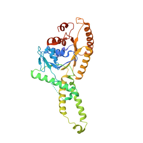
3A37

Structural insight into the membrane insertion of tail-anchored proteins by Get3


Experimental Data Snapshot

  • Method: X-RAY DIFFRACTION
  • Resolution: 3.00 Å
  • R-Value Free: 0.282 
  • R-Value Work: 0.253 
  • R-Value Observed: 0.253 

wwPDB Validation   3D Report Full Report


Ligand Structure Quality Assessment 


This is version 1.3 of the entry. See complete history


Literature

Structural insight into the membrane insertion of tail-anchored proteins by Get3

Yamagata, A.Mimura, H.Sato, Y.Yamashita, M.Yoshikawa, A.Fukai, S.

(2010) Genes Cells 15: 29-41

  • DOI: https://doi.org/10.1111/j.1365-2443.2009.01362.x
  • Primary Citation of Related Structures:  
    3A36, 3A37

  • PubMed Abstract: 

    Tail anchored (TA) proteins, which are important for numerous cellular processes, are defined by a single transmembrane domain (TMD) near the C-terminus. The membrane insertion of TA proteins is mediated by the highly conserved ATPase Get3. Here we report the crystal structures of Get3 in ADP-bound and nucleotide-free forms at 3.0 A and 2.8 A resolutions, respectively. Get3 consists of a nucleotide binding domain and a helical domain. Both structures exhibit a Zn(2+)-mediated homodimer in a head-to-head orientation, representing an open dimer conformation. Our cross-link experiments indicated the closed dimer-stimulating ATP hydrolysis, which might be coupled with TA-protein release. Further, our coexpression-based binding assays using a model TA protein Sec22p revealed the direct interaction between the helical domain of Get3 and the Sec22p TMD. This interaction is independent of ATP and dimer formation. Finally, we propose a structural mechanism that links ATP hydrolysis with the TA-protein insertion mediated by the conserved DTAPTGH motif.


  • Organizational Affiliation

    Structural Biology Laboratory, Life Science Division, Synchrotron Radiation Research Organization and Institute of Molecular and Cellular Biosciences, The University of Tokyo, Tokyo 113-0032, Japan.


Macromolecules
Find similar proteins by:  (by identity cutoff)  |  3D Structure
Entity ID: 1
MoleculeChains Sequence LengthOrganismDetailsImage
ATPase GET3
A, B
362Saccharomyces cerevisiaeMutation(s): 0 
Gene Names: 851458
EC: 3.6.3.16
UniProt
Find proteins for Q12154 (Saccharomyces cerevisiae (strain ATCC 204508 / S288c))
Explore Q12154 
Go to UniProtKB:  Q12154
Entity Groups  
Sequence Clusters30% Identity50% Identity70% Identity90% Identity95% Identity100% Identity
UniProt GroupQ12154
Sequence Annotations
Expand
  • Reference Sequence
Experimental Data & Validation

Experimental Data

  • Method: X-RAY DIFFRACTION
  • Resolution: 3.00 Å
  • R-Value Free: 0.282 
  • R-Value Work: 0.253 
  • R-Value Observed: 0.253 
  • Space Group: P 21 21 2
Unit Cell:
Length ( Å )Angle ( ˚ )
a = 115.229α = 90
b = 222.554β = 90
c = 49.291γ = 90
Software Package:
Software NamePurpose
CNSrefinement
HKL-2000data collection
HKL-2000data reduction
HKL-2000data scaling
CNSphasing

Structure Validation

View Full Validation Report


Ligand Structure Quality Assessment 


Entry History 

Deposition Data

Revision History  (Full details and data files)

  • Version 1.0: 2010-01-12
    Type: Initial release
  • Version 1.1: 2011-07-13
    Changes: Version format compliance
  • Version 1.2: 2018-02-14
    Changes: Experimental preparation
  • Version 1.3: 2024-03-13
    Changes: Data collection, Database references, Derived calculations