3ZBD

Structure of TGEV nsp1


Experimental Data Snapshot

  • Method: X-RAY DIFFRACTION
  • Resolution: 1.49 Å
  • R-Value Free: 0.209 
  • R-Value Work: 0.175 
  • R-Value Observed: 0.176 

wwPDB Validation   3D Report Full Report


This is version 1.1 of the entry. See complete history


Literature

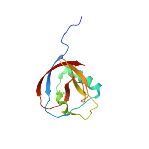
Structure of Alphacoronavirus Tgev Nsp1 Has Implications for Coronavirus Nsp1 Function and Evolution.

Jansson, A.M.

(2013) J Virol 87: 2949

  • DOI: https://doi.org/10.1128/JVI.03163-12
  • Primary Citation of Related Structures:  
    3ZBD

  • PubMed Abstract: 

    Coronavirus nsp1 has been shown to induce suppression of host gene expression and to interfere with the host immune response. However, the mechanism is currently unknown. The only available structural information on coronavirus nsp1 is the nuclear magnetic resonance (NMR) structure of the N-terminal domain of nsp1 from severe acute respiratory syndrome coronavirus (SARS-CoV) from the betacoronavirus genus. Here we present the first nsp1 structure from an alphacoronavirus, transmissible gastroenteritis virus (TGEV) nsp1. It displays a six-stranded β-barrel fold with a long alpha helix on the rim of the barrel, a fold shared with SARS-CoV nsp1(13-128). Contrary to previous speculation, the TGEV nsp1 structure suggests that coronavirus nsp1s have a common origin, despite the lack of sequence homology. However, comparisons of surface electrostatics, shape, and amino acid conservation between the alpha- and betacoronaviruses lead us to speculate that the mechanism for nsp1-induced suppression of host gene expression might be different in these two genera.


  • Organizational Affiliation

    Department of Cell and Molecular Biology, Uppsala University, Biomedical Center, Uppsala, Sweden. anna.jansson@icm.uu.se


Macromolecules
Find similar proteins by:  (by identity cutoff)  |  3D Structure
Entity ID: 1
MoleculeChains Sequence LengthOrganismDetailsImage
NON-STRUCTURAL PROTEIN 1
A, B
113Porcine transmissible gastroenteritis coronavirus strain PurdueMutation(s): 0 
UniProt
Find proteins for P0C6V2 (Porcine transmissible gastroenteritis coronavirus (strain Purdue))
Explore P0C6V2 
Go to UniProtKB:  P0C6V2
Entity Groups  
Sequence Clusters30% Identity50% Identity70% Identity90% Identity95% Identity100% Identity
UniProt GroupP0C6V2
Sequence Annotations
Expand
  • Reference Sequence
Experimental Data & Validation

Experimental Data

  • Method: X-RAY DIFFRACTION
  • Resolution: 1.49 Å
  • R-Value Free: 0.209 
  • R-Value Work: 0.175 
  • R-Value Observed: 0.176 
  • Space Group: P 1
Unit Cell:
Length ( Å )Angle ( ˚ )
a = 35.64α = 91.49
b = 36.09β = 108.85
c = 42.27γ = 94.2
Software Package:
Software NamePurpose
BUSTERrefinement
MOSFLMdata reduction
SCALAdata scaling

Structure Validation

View Full Validation Report


Entry History 

Deposition Data

Revision History  (Full details and data files)

  • Version 1.0: 2013-01-09
    Type: Initial release
  • Version 1.1: 2013-02-27
    Changes: Database references