3WX7

Crystal structure of COD


Experimental Data Snapshot

  • Method: X-RAY DIFFRACTION
  • Resolution: 1.35 Å
  • R-Value Free: 0.198 
  • R-Value Work: 0.174 
  • R-Value Observed: 0.175 

wwPDB Validation   3D Report Full Report


This is version 1.1 of the entry. See complete history


Literature

Structure-based analysis of domain function of chitin oligosaccharide deacetylase from Vibrio parahaemolyticus.

Hirano, T.Sugiyama, K.Sakaki, Y.Hakamata, W.Park, S.Y.Nishio, T.

(2015) FEBS Lett 589: 145-151

  • DOI: https://doi.org/10.1016/j.febslet.2014.11.039
  • Primary Citation of Related Structures:  
    3WX7

  • PubMed Abstract: 

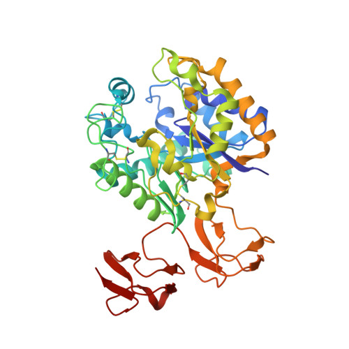
    The X-ray crystal structure of chitin oligosaccharide deacetylase from Vibrio parahaemolyticus (Vp-COD) was determined at an 1.35 Å resolution. The amino acid sequence and structure of Vp-COD show that the enzyme comprises one polysaccharide deacetylase domain (PDD) and two carbohydrate-binding domains (CBDs). On the basis of a chitin-binding assay with Vp-COD and its CBDs-deleted mutant, it was confirmed that CBDs can adhere to chitin. The catalytic activity of the CBDs-deleted mutant was only mildly depressed compared with that of Vp-COD, indicating that CBDs are unlikely to affect the configuration of the active center residues in active site of PDD.


  • Organizational Affiliation

    Department of Chemistry and Life Science, College of Bioresource Sciences, Nihon University, 1866 Kameino, Fujisawa, Kanagawa 252-0880, Japan.


Macromolecules
Find similar proteins by:  (by identity cutoff)  |  3D Structure
Entity ID: 1
MoleculeChains Sequence LengthOrganismDetailsImage
Chitin oligosaccharide deacetylase
A, B
402Vibrio parahaemolyticusMutation(s): 0 
Gene Names: cod1COD
UniProt
Find proteins for A6P4T5 (Vibrio parahaemolyticus)
Explore A6P4T5 
Go to UniProtKB:  A6P4T5
Entity Groups  
Sequence Clusters30% Identity50% Identity70% Identity90% Identity95% Identity100% Identity
UniProt GroupA6P4T5
Sequence Annotations
Expand
  • Reference Sequence
Small Molecules
Ligands 4 Unique
IDChains Name / Formula / InChI Key2D Diagram3D Interactions
GOL
Query on GOL

Download Ideal Coordinates CCD File 
F [auth A],
G [auth A],
K [auth B],
L [auth B]
GLYCEROL
C3 H8 O3
PEDCQBHIVMGVHV-UHFFFAOYSA-N
ZN
Query on ZN

Download Ideal Coordinates CCD File 
D [auth A],
I [auth B]
ZINC ION
Zn
PTFCDOFLOPIGGS-UHFFFAOYSA-N
ACT
Query on ACT

Download Ideal Coordinates CCD File 
E [auth A],
J [auth B]
ACETATE ION
C2 H3 O2
QTBSBXVTEAMEQO-UHFFFAOYSA-M
CA
Query on CA

Download Ideal Coordinates CCD File 
C [auth A],
H [auth B]
CALCIUM ION
Ca
BHPQYMZQTOCNFJ-UHFFFAOYSA-N
Experimental Data & Validation

Experimental Data

  • Method: X-RAY DIFFRACTION
  • Resolution: 1.35 Å
  • R-Value Free: 0.198 
  • R-Value Work: 0.174 
  • R-Value Observed: 0.175 
  • Space Group: P 21 21 21
Unit Cell:
Length ( Å )Angle ( ˚ )
a = 61.819α = 90
b = 104.106β = 90
c = 140.378γ = 90
Software Package:
Software NamePurpose
SCALEPACKdata scaling
SOLVEphasing
PHENIXrefinement
PDB_EXTRACTdata extraction
ADSCdata collection
HKL-2000data reduction
HKL-2000data scaling
PHENIXphasing

Structure Validation

View Full Validation Report


Entry History 

Deposition Data

Revision History  (Full details and data files)

  • Version 1.0: 2014-12-03
    Type: Initial release
  • Version 1.1: 2019-04-03
    Changes: Data collection, Database references