3WRU

Crystal structure of the bacterial ribosomal decoding site in complex with synthetic aminoglycoside with F-HABA group


Experimental Data Snapshot

  • Method: X-RAY DIFFRACTION
  • Resolution: 2.30 Å
  • R-Value Free: 0.262 
  • R-Value Work: 0.249 

wwPDB Validation   3D Report Full Report


Ligand Structure Quality Assessment 


This is version 1.1 of the entry. See complete history


Literature

Toxicity Modulation, Resistance Enzyme Evasion, and A-Site X-ray Structure of Broad-Spectrum Antibacterial Neomycin Analogs

Maianti, J.P.Kanazawa, H.Dozzo, P.Matias, R.D.Feeney, L.A.Armstrong, E.S.Hildebrandt, D.J.Kane, T.R.Gliedt, M.J.Goldblum, A.A.Linsell, M.S.Aggen, J.B.Kondo, J.Hanessian, S.

(2014) ACS Chem Biol 9: 2067-2073

  • DOI: https://doi.org/10.1021/cb5003416
  • Primary Citation of Related Structures:  
    3WRU

  • PubMed Abstract: 

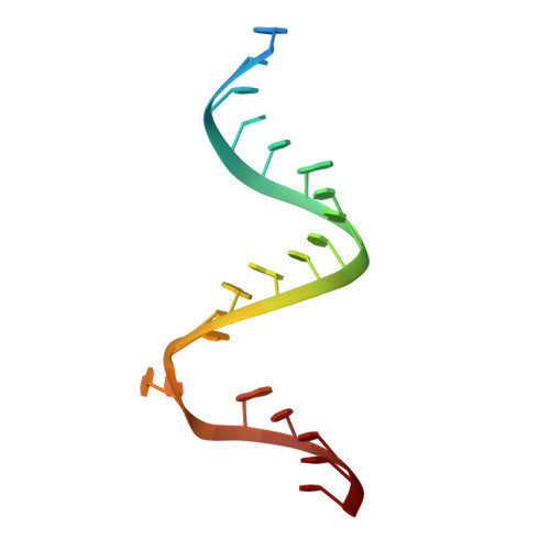
    Aminoglycoside antibiotics are pseudosaccharides decorated with ammonium groups that are critical for their potent broad-spectrum antibacterial activity. Despite over three decades of speculation whether or not modulation of pKa is a viable strategy to curtail aminoglycoside kidney toxicity, there is a lack of methods to systematically probe amine-RNA interactions and resultant cytotoxicity trends. This study reports the first series of potent aminoglycoside antibiotics harboring fluorinated N1-hydroxyaminobutyryl acyl (HABA) appendages for which fluorine-RNA contacts are revealed through an X-ray cocrystal structure within the RNA A-site. Cytotoxicity in kidney-derived cells was significantly reduced for the derivative featuring our novel β,β-difluoro-HABA group, which masks one net charge by lowering the pKa without compromising antibacterial potency. This novel side-chain assists in evasion of aminoglycoside-modifying enzymes, and it can be easily transferred to impart these properties onto any number of novel analogs.


  • Organizational Affiliation

    Department of Chemistry, Université de Montréal , CP 6128 Succ. Centre-Ville, Montréal, Québec H3C3J7, Canada.


Macromolecules

Find similar nucleic acids by:  Sequence   |   3D Structure  

Entity ID: 1
MoleculeChains LengthOrganismImage
RNA (5'-R(*UP*UP*GP*CP*GP*UP*CP*AP*CP*GP*CP*CP*GP*GP*CP*GP*AP*AP*GP*UP*CP*GP*C)-3')
A, B
23N/A
Sequence Annotations
Expand
  • Reference Sequence
Small Molecules
Ligands 2 Unique
IDChains Name / Formula / InChI Key2D Diagram3D Interactions
SJP
Query on SJP

Download Ideal Coordinates CCD File 
C [auth A],
D [auth B]
(2R,3R)-4-amino-N-[(1R,2S,3R,4R,5S)-5-amino-4-[(2,6-diamino-2,3,4,6-tetradeoxy-alpha-D-erythro-hexopyranosyl)oxy]-3-{[3-O-(2,6-diamino-2,3,4,6-tetradeoxy-beta-L-threo-hexopyranosyl)-beta-D-ribofuranosyl]oxy}-2-hydroxycyclohexyl]-3-fluoro-2-hydroxybutanamide
C27 H52 F N7 O11
XWTWBGQMVSXRIE-ZLDKHCKJSA-N
K
Query on K

Download Ideal Coordinates CCD File 
E [auth B]POTASSIUM ION
K
NPYPAHLBTDXSSS-UHFFFAOYSA-N
Experimental Data & Validation

Experimental Data

  • Method: X-RAY DIFFRACTION
  • Resolution: 2.30 Å
  • R-Value Free: 0.262 
  • R-Value Work: 0.249 
  • Space Group: P 1 21 1
Unit Cell:
Length ( Å )Angle ( ˚ )
a = 46.554α = 90
b = 32.602β = 108.5
c = 51.413γ = 90
Software Package:
Software NamePurpose
PHENIXmodel building
CNSrefinement
CrystalCleardata reduction
CrystalCleardata scaling
PHENIXphasing

Structure Validation

View Full Validation Report


Ligand Structure Quality Assessment 


Entry History 

Deposition Data

Revision History  (Full details and data files)

  • Version 1.0: 2014-11-05
    Type: Initial release
  • Version 1.1: 2024-03-20
    Changes: Data collection, Database references, Derived calculations