


Crystal Structure of Glycosyltransferase Vinc Involved in the Biosynthesis of Vicenistatin
Nango, E., Minami, A., Kumasaka, T., Eguchi, T.To be published.
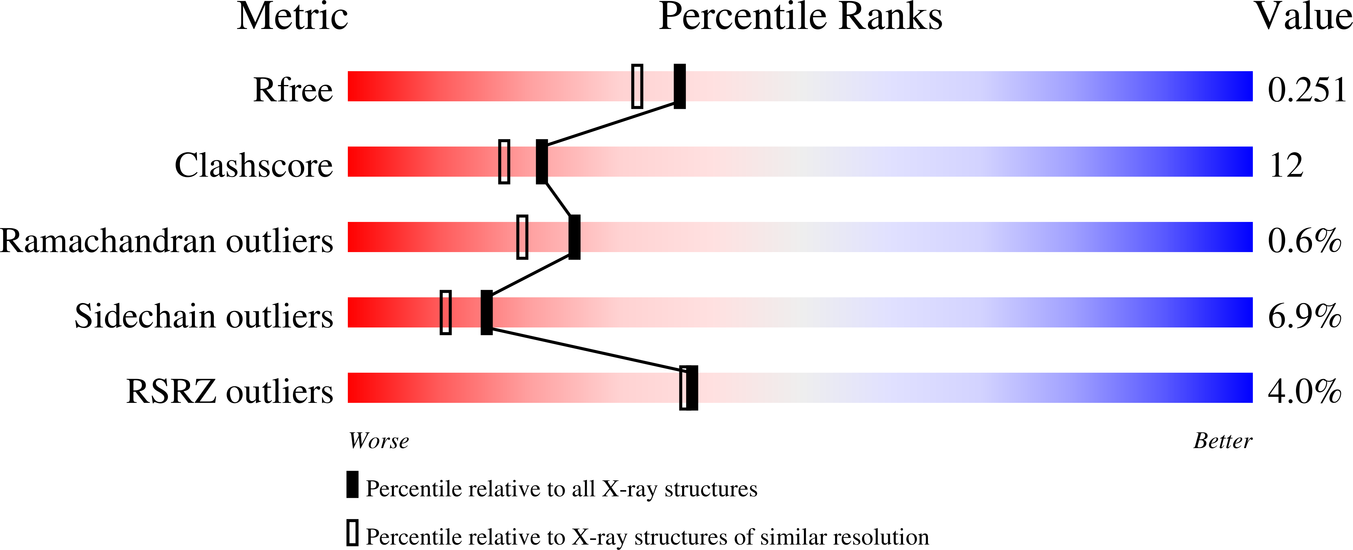
Experimental Data Snapshot
wwPDB Validation 3D Report Full Report
Entity ID: 1 | |||||
|---|---|---|---|---|---|
| Molecule | Chains | Sequence Length | Organism | Details | Image |
| Glycosyltransferase | 419 | Streptomyces halstedii | Mutation(s): 0 Gene Names: vinC | 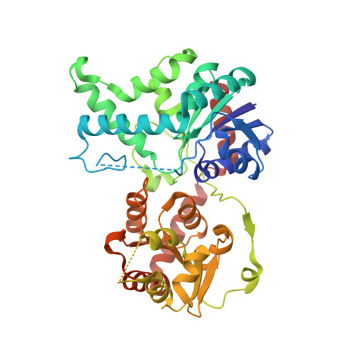 | |
UniProt | |||||
Find proteins for Q76KZ6 (Streptomyces halstedii) Explore Q76KZ6 Go to UniProtKB: Q76KZ6 | |||||
Entity Groups | |||||
| Sequence Clusters | 30% Identity50% Identity70% Identity90% Identity95% Identity100% Identity | ||||
| UniProt Group | Q76KZ6 | ||||
Sequence AnnotationsExpand | |||||
| |||||
| Ligands 1 Unique | |||||
|---|---|---|---|---|---|
| ID | Chains | Name / Formula / InChI Key | 2D Diagram | 3D Interactions | |
| MG Query on MG | C [auth A] | MAGNESIUM ION Mg JLVVSXFLKOJNIY-UHFFFAOYSA-N |  | ||
| Length ( Å ) | Angle ( ˚ ) |
|---|---|
| a = 98.205 | α = 90 |
| b = 130.392 | β = 90 |
| c = 140.107 | γ = 90 |
| Software Name | Purpose |
|---|---|
| BSS | data collection |
| SOLVE | phasing |
| REFMAC | refinement |
| HKL-2000 | data reduction |
| HKL-2000 | data scaling |
RCSB PDB (citation) is hosted by
RCSB PDB is a member of the

